
Page 467 - Atlas of Histology with Functional Correlations
P. 467
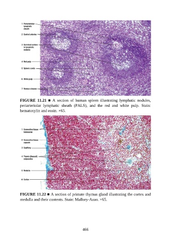
FIGURE  11.21  ■  A  section  of  human  spleen  illustrating  lymphatic  nodules,
               periarteriolar  lymphatic  sheath  (PALS),  and  the  red  and  white  pulp.  Stain:
               hematoxylin and eosin. ×65.


               FIGURE 11.22 ■ A section of primate thymus gland illustrating the cortex and
               medulla and their contents. Stain: Mallory-Azan. ×65.


                                                          466