
Page 581 - Atlas of Histology with Functional Correlations
P. 581
ADDITIONAL HISTOLOGIC IMAGES
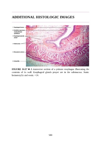
FIGURE 14.17 ■ A transverse section of a primate esophagus illustrating the
contents of its wall. Esophageal glands proper are in the submucosa. Stain:
hematoxylin and eosin. ×10.
580