
Page 697 - Atlas of Histology with Functional Correlations
P. 697
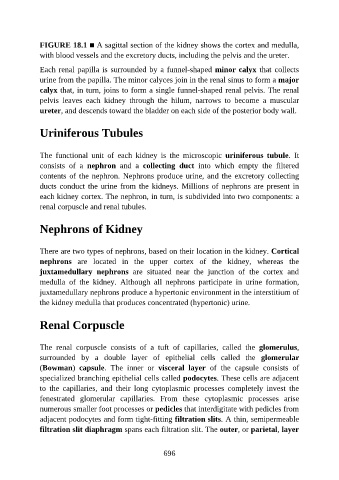
FIGURE 18.1 ■ A sagittal section of the kidney shows the cortex and medulla,

               with blood vessels and the excretory ducts, including the pelvis and the ureter.

               Each renal papilla is surrounded by a funnel-shaped minor calyx that collects
               urine from the papilla. The minor calyces join in the renal sinus to form a major
               calyx that, in turn, joins to form a single funnel-shaped renal pelvis. The renal
               pelvis  leaves  each  kidney  through  the  hilum,  narrows  to  become  a  muscular

               ureter, and descends toward the bladder on each side of the posterior body wall.


               Uriniferous Tubules


               The  functional  unit  of  each  kidney  is  the  microscopic  uriniferous  tubule.  It

               consists  of  a  nephron  and  a  collecting  duct  into  which  empty  the  filtered
               contents of the nephron. Nephrons produce urine, and the excretory collecting
               ducts conduct the urine from the kidneys. Millions of nephrons are present in
               each kidney cortex. The nephron, in turn, is subdivided into two components: a

               renal corpuscle and renal tubules.


               Nephrons of Kidney


               There are two types of nephrons, based on their location in the kidney. Cortical

               nephrons  are  located  in  the  upper  cortex  of  the  kidney,  whereas  the
               juxtamedullary  nephrons  are  situated  near  the  junction  of  the  cortex  and
               medulla  of  the  kidney.  Although  all  nephrons  participate  in  urine  formation,
               juxtamedullary nephrons produce a hypertonic environment in the interstitium of

               the kidney medulla that produces concentrated (hypertonic) urine.


               Renal Corpuscle


               The  renal  corpuscle  consists  of  a  tuft  of  capillaries,  called  the  glomerulus,
               surrounded  by  a  double  layer  of  epithelial  cells  called  the  glomerular
               (Bowman)  capsule.  The  inner  or  visceral  layer  of  the  capsule  consists  of
               specialized branching epithelial cells called podocytes. These cells are adjacent
               to  the  capillaries,  and  their  long  cytoplasmic  processes  completely  invest  the

               fenestrated  glomerular  capillaries.  From  these  cytoplasmic  processes  arise
               numerous smaller foot processes or pedicles that interdigitate with pedicles from
               adjacent podocytes and form tight-fitting filtration slits. A thin, semipermeable

               filtration slit diaphragm spans each filtration slit. The outer, or parietal, layer


                                                          696