
Page 791 - Atlas of Histology with Functional Correlations
P. 791
A thick connective tissue capsule, the tunica albuginea, surrounds each testis
(Fig. 20.1). On the posterior side, the tunica albuginea thickens and extends
inward into each testis to form the mediastinum testis. A thin connective tissue
septum extends from the mediastinum testis and subdivides each testis into
about 250 incomplete compartments or testicular lobules, each containing one
to four highly coiled seminiferous tubules. Each seminiferous tubule is lined
with a stratified germinal epithelium, containing proliferating spermatogenic
(germ) cells and nonproliferating supporting (sustentacular), or Sertoli, cells.
The seminiferous tubules are the site of spermatogenic cell division, maturation,
and transformation into sperm.
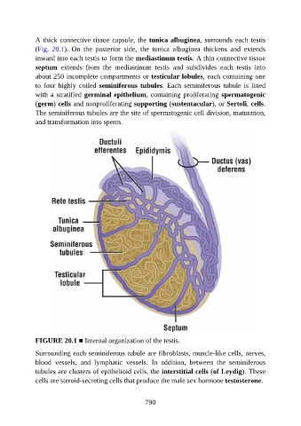
FIGURE 20.1 ■ Internal organization of the testis.
Surrounding each seminiferous tubule are fibroblasts, muscle-like cells, nerves,
blood vessels, and lymphatic vessels. In addition, between the seminiferous
tubules are clusters of epithelioid cells, the interstitial cells (of Leydig). These
cells are steroid-secreting cells that produce the male sex hormone testosterone.
790