
Page 786 - Atlas of Histology with Functional Correlations
P. 786
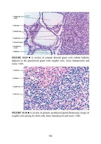
FIGURE  19.19  ■  A  section  of  primate  thyroid  gland  with  colloid  follicles

               adjacent  to  the  parathyroid  gland  with  oxyphil  cells.  Stain:  hematoxylin  and
               eosin. ×100.


               FIGURE 19.20 ■ A section of primate parathyroid gland illustrating clumps of
               oxyphil cells among the chief cells. Stain: hematoxylin and eosin. ×100.


                                                          785