
Page 785 - Atlas of Histology with Functional Correlations
P. 785
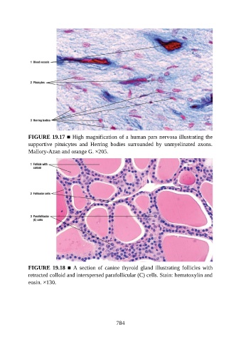
FIGURE 19.17 ■ High magnification of a human pars nervosa illustrating the
               supportive  pituicytes  and  Herring  bodies  surrounded  by  unmyelinated  axons.
               Mallory-Azan and orange G. ×205.


               FIGURE 19.18 ■ A section of canine thyroid gland illustrating follicles with
               retracted colloid and interspersed parafollicular (C) cells. Stain: hematoxylin and
               eosin. ×130.


                                                          784