
Page 834 - Atlas of Histology with Functional Correlations
P. 834
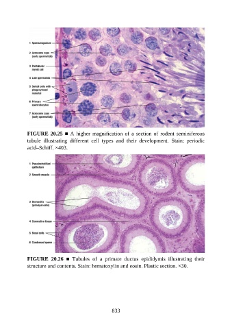
FIGURE 20.25 ■ A higher magnification of a section of rodent seminiferous
               tubule  illustrating  different  cell  types  and  their  development.  Stain:  periodic

               acid–Schiff. ×403.


               FIGURE  20.26  ■  Tubules  of  a  primate  ductus  epididymis  illustrating  their
               structure and contents. Stain: hematoxylin and eosin. Plastic section. ×30.


                                                          833