
Page 434 - AWSAR 2.0
P. 434
410 || AWSAR Awarded Popular Science Stories - 2019
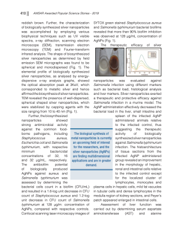
reddish brown. Further, the characterization of biologically synthesized silver nanoparticles was accomplished by employing various biophysical techniques such as UV visible spectra, x-ray diffraction, scanning electron microscope (SEM), transmission electron microscopy (TEM) and Fourier-transform infrared analysis. The shape of biosynthesized silver nanoparticles as determined by field emission SEM micrographs was found to be spherical and monodispersed (Fig. 1). The elemental profile of biologically synthesized silver nanoparticles, as analysed by energy- dispersive x-ray analysis graphs, showed the optical absorption peak at 3KeV, which corresponded to metallic silver and hence affirmed the biosynthesis of silver nanoparticles. TEM revealed the presence of well-dispersed, spherical shaped silver nanoparticles, which were stabilized by capping agents with the size ranging from 10 to 40 nm (Fig. 1).
Further, the biosynthesized
nanoparticles showed
strong antimicrobial activity
against the common food-
borne pathogens, including Staphylococcus aureus,
Escherichia coli and Salmonella
typhimurium, with respective
minimum bactericidal concentrations of 32, 16
and 32 μg/mL, respectively.
The antibiofilm potential
of biologically produced
AgNPs against aureus and
Salmonella typhimurium was
assessed by determining the
bacterial cells count in a biofilm (CFU/mL) and resulted in a 1.6-log unit decrease in CFU count of Staphylococcus aureus and 1.2-log unit decrease in CFU count of Salmonella typhimurium at 128 μg/m: concentration of AgNPs, compared with respective controls. Confocal scanning laser microscopy images of
SYTOX green stained Staphylococcus aureus and Salmonella typhimurium bacterial biofilms revealedthatmorethan90%biofilminhibition was observed at 128 μg/mL concentration of AgNPs (Fig. 1).
The therapeutic efficacy of silver
nanoparticles was evaluated against Salmonella infection using different markers such as bacterial load, histological analysis and liver markers. Silver nanoparticles exerted a therapeutic and protective efficacy against Salmonella infection in a murine model. The AgNP administration effectively decreased the bacterial load in the liver, small intestine and
spleen of the infected AgNP administered animals relative to the infected control, thus suggesting the therapeutic activity of biologically synthesized silver nanoparticles against Salmonella typhimurium infection. The histoarchitecture of tissue sections from the infected AgNP administered group revealed an improvement in the morphology of hepatic, renal and intestinal cells relative to the infected control except for the localized cluster of lymphocytes, monocytes and
plasma cells in hepatic cells, mild fat vacuoles in tubular cells and dense lymphocytes in the medulla region of kidney sections, and Peyer’s patch appeared enlarged in intestinal cells.
Assessment of liver function was carried out by determining serum aspartate aminotransferase (AST) and alanine
The biological synthesis of metal nanoparticles is currently an upcoming field of interest for the researchers, and the silver nanoparticles (AgNPs) are finding multidimensional applications and are in greater demand.
