
Page 50 - CJO_F18
P. 50
C CLINICAL RESEARCH
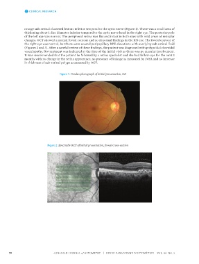
orange sub-retinal clustered lesions inferior temporal to the optic nerve (Figure 1). There was a small area of
thickening about 1 disc diameter inferior temporal to the optic nerve head in the right eye. The posterior pole
of the left eye was normal. The peripheral retina was flat and intact in both eyes with mild areas of reticular
changes. OCT showed a normal foveal contour and no abnormal findings in the left eye. The foveal contour of
the right eye was normal, but there were several peripapillary RPE elevations with overlying sub-retinal fluid
(Figures 2 and 3). After a careful review of these findings, the patient was diagnosed with polypoidal choroidal
vasculopathy. No treatment was indicated at the time of the initial visit as there was no macular involvement.
It was recommended that the patient be followed by a retina specialist and she had follow-ups for the next 3
months with no change in the retina appearance, no presence of leakage as measured by IVFA and no increase
in thickness of sub-retinal polyps as assessed by OCT.
188 Photos
Figure 1: Fundus photograph of initial presentation, OD
Photos
188
189
190 Figure 1: Fundus photograph of initial presentation, OD
189
Figure 2: Spectralis OCT of initial presentation, foveal cross-section
190 Figure 1: Fundus photograph of initial presentation, OD
191
192 Figure 2: Spectralis OCT of initial presentation, foveal cross-section
191
192 Figure 2: Spectralis OCT of initial presentation, foveal cross-section
50 CANADIAN JOURNAL of OPTOMETRY | REVUE CANADIENNE D’OPTOMÉTRIE VOL. 80 NO. 3
38668_CJO_F18 August 10, 2018 8:58 AM APPROVAL: ___________________ DATE: ___________________