
Page 53 - CJO_F18
P. 53
CASE REPORT
202
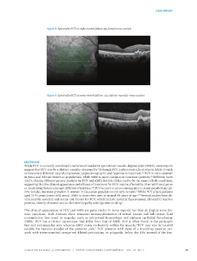
Figure 8: Spectralis OCT at eight-month follow-up, foveal cross-section
203 Figure 7: Fundus photograph at seven-month follow-up
204
205 Figure 8: Spectralis OCT at eight-month follow-up, foveal cross-section
Figure 9: Spectralis OCT at seven month follow- up, inferior macular cross-section
206
207 Figure 9: Spectralis OCT at seven month follow- up, inferior macular cross-section
DISCUSSION 208
While PCV is currently considered a variation of exudative age-related macular degeneration (AMD), some reports
209
suggest that PCV may be a distinct vascular abnormality. Although PCV is phenotypically similar to AMD, it tends
2
210
to have a very different natural progression, target demographic and response to treatment. PCV is more common
2,6
in Asian and African-American populations, while AMD is more common in Caucasian patients. Different races
2,6
tend to display different genetic markers for PCV and AMD, but risk alleles tend to be the same in both conditions,
suggesting that the clinical appearance and efficacy of treatment for PCV may be affected by other additional genes
or modulating factors amongst different ethnicities. 1,2,6 PCV is more common among men in Asian populations (22-
37% female), but more prevalent in women in Caucasian populations (52-65% female). While PCV affects patients
2
aged 21-93 years (mean 68.4 years), AMD is most often seen at around 80 years of age. 2,6,7 Several studies have de-
termined the systemic and ocular risk factors for PCV, which include systemic hypertension, elevated C-reactive
proteins, history of central serous chorioretinopathy and cigarette smoking. 2
The clinical appearances of PCV and AMD are quite similar in some regards, but they do display some dis-
tinct variations. Both diseases show abnormal neovascularization of retinal tissues and sub-retinal fluid
accumulation that result in sequelae such as sub-retinal hemorrhage and pigment epithelial detachment
(PED). PCV has a clinical appearance that differ from that of AMD. PCV is often found in the peripapil-
1
lary and extramacular area whereas AMD exists exclusively within the macula. PCV may also be located
2
outside the vascular arcades of the posterior pole. PCV presents with signs of a branching vascular net-
2
work with interconnected orange-red dilated protrusions, or polypoids, below the RPE instead of the dru-
CANADIAN JOURNAL of OPTOMETRY | REVUE CANADIENNE D’OPTOMÉTRIE VOL. 80 NO. 3 53
38668_CJO_F18 August 10, 2018 8:58 AM APPROVAL: ___________________ DATE: ___________________