
Page 51 - CJO_F18
P. 51
CASE REPORT
Figure 3: Spectralis OCT of initial presentation, inferior macular cross-section
193
194 Figure 3: Spectralis OCT of initial presentation, inferior macular cross-section
Approximately 4 months after the diagnosis, the patient presented at the clinic for an emergency visit with complaints of
195
blurred vision and new onset of flashes and floaters in the right eye for two days. Her vision with glasses was 20/40-1 with
eccentric viewing OD and 20/20 with the left eye. There was no improvement with refraction. Her pupils were equal,
round, and reactive to light, and there was no APD. Ocular motilities were full and smooth OD, OS and her confrontation
visual fields were full with finger-counting in all quadrants in both eyes. The right eye had central metamorphopsia and
an incomplete central scotoma as measured on an Amsler grid. Anterior slit lamp examination showed no remarkable
findings. Goldmann applanation tonometry measured pressures of 17mmHg in the right eye and 19mmHg in the left eye.
Dilated fundus exam showed trace nuclear sclerosis of the lens in both eyes and a posterior vitreous detachment in both
eyes. The optic nerves had a C/D ratio of 0.5 round in both eyes, and were pink and distinct. The a/v ratio was 2/3 with
normal caliber and no crossing changes were noted in either eye. The macula and posterior pole of the left eye were flat
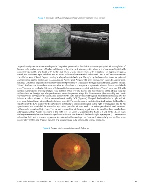
without fluid. In the right eye, a large sub-retinal hemorrhage measuring 5 disc diameters (DD) horizontal by 4DD verti-
cal was present throughout the macula and inferior to the optic nerve with overlying sub-retinal fluid extending into the
inferior arcade as viewed on clinical exam and confirmed by OCT (Figure 4). The peripheries of both the right and left
eyes were flat and intact without breaks, holes or tears. OCT showed a large area of significant sub-retinal fluid and large
elevations in the RPE inferior to the optic nerve extending to the macular region in the right eye (Figures 5 and 6). An
appointment was scheduled for evaluation by a retina specialist within a week. The retina specialist initiated treatment
with Avastin intravitreal injections. The patient returned for a follow-up appointment in our clinic four months later,
after receiving four Avastin injections in the right eye. Her vision was corrected to 20/30 OD and 20/20 OS. All exam
findings were stable, but she showed a significant reduction in sub-retinal fluid in the right eye (Figure 7). There was no
sub-retinal fluid in the macular region and the sub-retinal hemorrhage had decreased substantially to a small area ap-
proximately 1DD in size (Figures 8 and 9). She has continued to be followed by a retina specialist.
196
197 Figure 4: Fundus photograph at four-month follow up
193
194 Figure 4: Fundus photograph at four-month follow up
Figure 3: Spectralis OCT of initial presentation, inferior macular cross-section
195
196
197 Figure 4: Fundus photograph at four-month follow up
CANADIAN JOURNAL of OPTOMETRY | REVUE CANADIENNE D’OPTOMÉTRIE VOL. 80 NO. 3 51
38668_CJO_F18 August 10, 2018 8:58 AM APPROVAL: ___________________ DATE: ___________________