
Page 26 - CJO_W18
P. 26
C CLINICAL RESEARCH
Slit lamp examination showed mild bilateral blepharitis, clear conjunctiva, and clear cornea without endothelial
pigment or keratic precipitates. The anterior chamber was deep and quiet by Van Herick angle estimation and the
iris was normal without signs of atrophy, obvious posterior synechiae, or trans-illumination defects. Intraocular
pressures (IOP) were 17 OD, 15 OS at 08:01 by Goldmann applanation tonometry.
Dilated examination showed trace nuclear sclerosis and cortical opacities without evidence of pseudoexfoliation or
pigment. The macula, vessels, and periphery were all normal OD, OS. There was a posterior vitreous detachment
OD, OS with no evidence of peripheral retinal abnormality.
The optic nerve head was average to large size OD>OS based on the vertical disc height using the adjusted slit
lamp graticule and a Volk 78 D lens with a correction factor of 1.2x. The optic cup was of moderate depth, with
early visible laminar dots OU. There was mild alpha zone parapapillary atrophy, but no signs of pallor or disc
hemorrhages OU. There was a subtle inferior retinal nerve fiber layer wedge defect with associated inferior
rim thinning, inferior vessel baring, and inferior arteriole narrowing OU. Additionally, the superior rim was
suspicious for glaucomatous optic neuropathy OU with evidence of early vessel baring OD>OS and relative
thinning compared to other optic nerve sectors. Cup-to-disc ratios were estimated to be 0.7 v/0.7 h OD and
0.75 v/0.7 h OS.
Baseline photos and optical coherence tomography (OCT) Optic Nerve Head (ONH) and Retinal Nerve Fiber Layer
(RNFL) Analysis were acquired. Both subjective and objective imaging confirmed the findings in the clinical exam,
as shown in Figures 1 and 2.
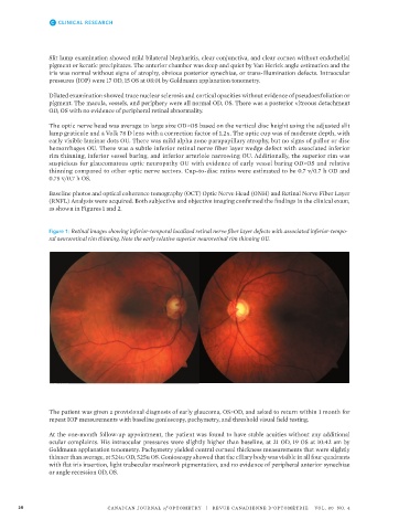
Figure 1: Retinal images showing inferior-temporal localized retinal nerve fiber layer defects with associated inferior-tempo-
ral neuroretinal rim thinning. Note the early relative superior neuroretinal rim thinning OU.
Figure 1: Retinal images showing inferior-temporal localized retinal nerve fiber
layer defects with associated inferior-temporal neuroretinal rim thinning. Note
The patient was given a provisional diagnosis of early glaucoma, OS>OD, and asked to return within 1 month for
repeat IOP measurements with baseline gonioscopy, pachymetry, and threshold visual field testing.
the early relative superior neuroretinal rim thinning OU.
At the one-month follow-up appointment, the patient was found to have stable acuities without any additional
ocular complaints. His intraocular pressures were slightly higher than baseline, at 21 OD, 19 OS at 10:42 am by
Goldmann applanation tonometry. Pachymetry yielded central corneal thickness measurements that were slightly
thinner than average, at 524u OD, 525u OS. Gonioscopy showed that the ciliary body was visible in all four quadrants
with flat iris insertion, light trabecular meshwork pigmentation, and no evidence of peripheral anterior synechiae
or angle recession OD, OS.
26 CANADIAN JOURNAL of OPTOMETRY | REVUE CANADIENNE D’OPTOMÉTRIE VOL. 80 NO. 4
5