
Page 27 - CJO_W18
P. 27
CASE STUDY
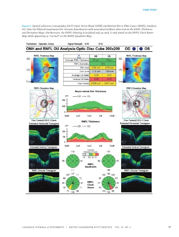
Figure 2: Optical coherence tomography (OCT) Optic Nerve Head (ONH) and Retinal Nerve Fiber Layer (RNFL) Analysis
OU. Note the bilateral nasal posterior vitreous detachments with associated artifacts observed on the RNFL Thickness
and Deviation Maps. Furthermore, the RNFL thinning is localized and, as such, is only noted on the RNFL Clock Hours
Map while appearing as “normal” on the RNFL Quadrant Map.
Figure 2: Optical coherence tomography (OCT) Optic Nerve Head (ONH) and
Retinal Nerve Fiber Layer (RNFL) Analysis OU. Note the bilateral nasal posterior
vitreous detachments with associated artifacts observed on the RNFL Thickness
CANADIAN JOURNAL of OPTOMETRY | REVUE CANADIENNE D’OPTOMÉTRIE VOL. 80 NO. 4 27
and Deviation Maps. Furthermore, the RNFL thinning is localized and, as such, is
6