
Page 28 - CJO_W18
P. 28
C CLINICAL RESEARCH
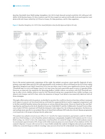
Baseline threshold visual field testing (Humphrey 24-2 SITA Fast) showed normal-sensitivity OD with good reli-
ability (0/10 fixation losses, 5% false positives, and 0% false negatives) and several focally depressed superior nasal
defects OS with lower reliability (5/11 fixation losses, 3% false positives, and 8% false negatives).
Figure 3: Baseline Humphrey 24-2 SITA Fast visual field plots show focally depressed defects OS only.
Due to the normal gonioscopic appearance of the angle, the patient was given a more specific diagnosis of early
primary open angle glaucoma OS>OD with questionable early perimetric defects OS based on the SITA Fast testing
algorithm. Bengtsson and Heijl found that SITA Fast test times (avg 5.0 min) were significantly shorter than Full
4
Threshold (avg 14.6 min) and Fastpac (avg 9.4 min) test times, but were essentially equal in terms of reproducibility.
However, as expected, the sensitivity for detecting shallow (subtle) defects was greater with Full Threshold test-
ing. In other words, shallow defects noted in Full Threshold testing were progressively less visible (perhaps even
absent) with Fastpac and SITA Fast, while the detection of deeper focal defects was essentially identical with all
three strategies.
Through collaboration with the patient, we decided to monitor the condition closely at that time without treatment
until repeat structural and functional testing confirmed the suspected defects (and/or suggested progression) and
to further establish baseline intraocular pressures at various diurnal time points. However, based on the case find-
ings thus far, and based on the Early Manifest Glaucoma Trial (EMGT) and the Collaborative Normal Tension Glau-
coma Study (CNTGS), treatment will likely be initiated to help reduce the risk of disease progression. Furthermore,
and perhaps more specific to our patient, Kim et al. found that sufficient IOP reduction slowed disease progression
even in patients with suspected preperimetric glaucoma. 5
The patient was asked to return to our office in 3-4 months for repeat tonometry, pachymetry, and 24-2 thresh-
old visual field testing, as well as baseline optical coherence tomography Ganglion Cell Analysis and 10-2 visual
field testing. As a brief review, the importance (and benefits) of central 10-degree visual field analysis cannot
be overstated: approximately 50% of eyes with mild-moderate glaucoma were found to have defects within the
central 3 degrees, 11 eyes with normal 24-2 visual fields outside the central 10 degrees showed arcuate defects
6
within the central 10 degrees with 10-2 testing, nine percent of normal 30-2 threshold visual fields in glaucoma
7
28 CANADIAN JOURNAL of OPTOMETRY | REVUE CANADIENNE D’OPTOMÉTRIE VOL. 80 NO. 4
8