
Page 43 - CJO_W18
P. 43
CASE STUDY
sensitivity of several points and worsened mean and pattern standard deviation, with a few scattered superonasal
rim defects in the right eye (Figure 2); the left eye showed an incomplete inferior arcuate defect with a cluster of
defects superonasally, which was slightly improved in both hemifields (Figure 3). The retinal nerve fiber layer by
optical coherence tomography was stable in the right eye, and still grossly elevated, but with an average thickness
of -31 μm (123 μm) in the left eye compared to previously.
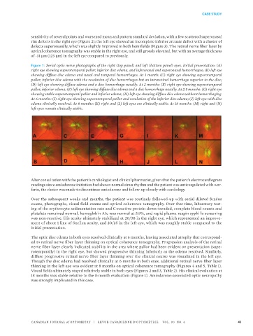
Figure 1: Serial optic nerve photographs of the right (top panel) and left (bottom panel) eyes. Initial presentation: (A)
right eye showing superotemporal pallor, inferior disc edema, and inferonasal and superonasal hemorrhages; (B) left eye
showing diffuse disc edema and nasal and temporal hemorrhages. At 1 month: (C) right eye showing superotemporal
pallor, inferior disc edema with the resolution of disc hemorrhages but an intraretinal hemorrhage superior to the disc;
(D) left eye showing diffuse edema and a disc hemorrhage nasally. At 2 months: (E) right eye showing superotemporal
pallor, inferior edema; (F) left eye showing diffuse disc edema and a disc hemorrhage nasally. At 2.5 months: (G) right eye
showing stable superotemporal pallor and inferior edema; (H) left eye showing diffuse disc edema without hemorrhaging.
At 6 months: (I) right eye showing superotemporal pallor and resolution of the inferior disc edema; (J) left eye with disc
edema clinically resolved. At 8 months: (K) right and (L) left eyes are clinically stable. At 18 months: (M) right and (N)
left eyes remain clinically stable.
After consultation with the patient’s cardiologist and clinical pharmacist, given that the patient’s electrocardiogram
readings since amiodarone initiation had shown normal sinus rhythm and the patient was anticoagulated with war-
farin, the choice was made to discontinue amiodarone and follow-up closely with cardiology.
Over the subsequent weeks and months, the patient was routinely followed-up with serial dilated fundus
exams, photographs, visual field exams and optical coherence tomography. Over that time, laboratory test-
ing of the erythrocyte sedimentation rate and C-reactive protein down-trended, complete blood counts and
platelets remained normal, hemoglobin A1c was normal at 5.9%, and rapid plasma reagin syphilis screening
was non-reactive. His acuity ultimately stabilized at 20/30 in the right eye, which represented an improve-
ment of about 1 line of Snellen acuity, and 20/25 in the left eye, which was roughly stable compared to the
initial presentation.
The optic disc edema in both eyes resolved clinically at 6 months, leaving associated atrophy that correspond-
ed to retinal nerve fiber layer thinning on optical coherence tomography. Progression analysis of the retinal
nerve fiber layer clearly indicated stability in the area where pallor had been evident on presentation (supe-
rotemporally) in the right eye, but showed progressive thinning inferiorly as the edema resolved. Similarly,
diffuse progressive retinal nerve fiber layer thinning over the clinical course was visualized in the left eye.
Though the disc edema had resolved clinically at 6 months in both eyes, additional retinal nerve fiber layer
thinning in the left eye was evident at 8 months on optical coherence tomography (Figures 4 and 5, Table 1).
Visual fields ultimately stayed relatively stable in both eyes (Figures 2 and 3, Table 2). His clinical evaluation at
18 months was stable relative to the 8-month evaluation (Figure 1). Amiodarone-associated optic neuropathy
was strongly implicated in this case.
CANADIAN JOURNAL of OPTOMETRY | REVUE CANADIENNE D’OPTOMÉTRIE VOL. 80 NO. 4 43