
Page 47 - CJO_W18
P. 47
CASE STUDY
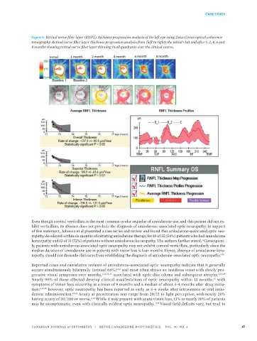
Figure 5: Retinal nerve fiber layer (RNFL) thickness progression analysis of the left eye using Zeiss Cirrus optical coherence
tomography. Retinal nerve fiber layer thickness progression analysis from (left to right) the initial visit and after 1, 2, 4, 6 and
8 months showing retinal nerve fiber layer thinning in all quadrants over the clinical course.
Even though corneal verticillata is the most common ocular sequelae of amiodarone use, and this patient did not ex-
hibit verticillata, its absence does not preclude the diagnosis of amiodarone-associated optic neuropathy. In support
of this statement, Johnson et al presented a case series and review and found that amiodarone-associated optic neu-
ropathy developed within six months of initiating amiodarone therapy for 19 of 35 (54%) patients who had amiodarone
keratopathy and 13 of 18 (72%) of patients without amiodarone keratopathy. The authors further stated, “Consequent-
ly, patients with amiodarone-associated optic neuropathy may not exhibit corneal verticillate, particularly since the
median duration of amiodarone use in patients with vision loss is four months. Hence, absence of amiodarone kera-
topathy should not dissuade clinicians from establishing the diagnosis of amiodarone-associated optic neuropathy.” 21
Reported cases and cumulative reviews of amiodarone-associated optic neuropathy indicate that it generally
occurs simultaneously bilaterally (around 66%) 2,22 and most often shows an insidious onset with slowly pro-
gressive visual symptoms over months, 2,18,22,23 associated with optic disc edema and subsequent atrophy. 2,23,24
Nearly 90% of those affected develop clinical manifestations of optic neuropathy within 12 months, with
18
symptoms of vision loss occurring at a mean of 9 months and a median of about 3-4 months after drug initia-
tion; 2,18,25 however, optic neuropathy has been reported as early as 3-4 weeks after intravenous or oral amio-
darone administration. 13,14 Acuity at presentation may range from 20/15 to light perception, with nearly 20%
having acuity of 20/200 or worse. 2,18 While it may present with acute vision loss, 13% to nearly 30% of patients
may be asymptomatic, even with clinically evident optic neuropathy. 2,18 Visual field deficits vary, but tend to
CANADIAN JOURNAL of OPTOMETRY | REVUE CANADIENNE D’OPTOMÉTRIE VOL. 80 NO. 4 47