
Page 11 - CJO_W17
P. 11
CASE STUDY
This case report describes a symptomatic patient who was frustrated and unclear as to the source of her unstable
vision and discomfort, which had several etiologies, including DE and corneal dystrophy.
CASE REPORT
A 62-year-old Caucasian female was referred to a DE clinic due to longstanding symptoms of fluctuating vision
and DE. Her most recent eye exam was 5 months prior; a refractive change was noted and new glasses were pre-
scribed. Her general health revealed a history of fibromyalgia, rheumatoid arthritis, hypertension, hypothyroidism
and depression. Medication use included Diovan HCT (Novartis) for her hypertension, Synthroid (Abbvie) for
®
®
her thyroid, Xanax (Pfizer) for her depression, vitamins (E and C) and omega-3 supplements. Her ocular history
®
revealed longstanding complaints of DE, fluctuating vision, pain, irritation and a gritty sensation in both eyes. The
patient also reported dryness of the mouth, throat, and nose; she tested negative for Sjögren’s syndrome. Addition-
ally, macular drusen were noted and age-related macular degeneration (ARMD) was diagnosed, for which she takes
vitamins (Vitalux , ALCON) and is being followed by a retinal specialist. Mild nuclear sclerosis (grade 1) was noted
®
in both eyes. The patient reported using artificial tears (Systane ULTRA, ALCON) 8X/day and an ocular ointment
®
at bedtime (Liposic gel, Bausch + Lomb) to address her ocular discomfort. She remained unsatisfied with her vision
®
despite her new glasses and continued to report ocular discomfort.
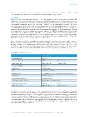
A comprehensive DE evaluation revealed severe symptoms (score of 87.5/100) using the Ocular Surface Disease In-
dex (OSDI) questionnaire. Other tear tests were performed and the results are summarized in Table 2; most values
were within normal limits. Distance acuities were similar to those reported at the annual eye exam (OD 6/7.5 ; OS
+2
6/6 ; OU 6/6 ). The near acuity revealed less than optimal results (OD 0.8 M; OS 1.0 M at 40 cm using a near point
-1
+2
card), which differed from the results at her check-up 5 months previously (OD 0.37; OS 0.50 M at 50 cm).
Table 2: Clinical findings of DE exam
Tests Clinical Findings
OSDI questionnaire 87.5/100
Osmolarity (TearLab) OD 288 mOsml/L OS 291 mOsml/L
Cotton Thread Test OD 34 mm/15 sec OS 36 mm/15 sec
Tear meniscus height 0.2 mm OU
Eyelid margin Telangiectasia OU
Lashes Cylindrical dandruff OU
Meibomian glands Yellow, liquid secretions, non-linear with missing glands OU
Corneal staining No defects noted OU
Bulbar Conjunctival Staining No defects noted OU
Palpebral Conjunctival Staining ULMS <20% along the full length, OU
Tear Break Up Time (TBUT) OD 3 sec OS 2 sec
Endothelial cell count OD 2367 cells/mm 2 OS 2423 cells/mm 2
A detailed anterior segment evaluation revealed redness (telangiectasia) along the eyelid margin, clear gelati-
nous deposits at the base of a few lashes resembling cylindrical dandruff (CD) and several differently shaped
translucent corneal opacities in both eyes. Epilation was performed on the lashes that had CD and a micro-
scopic evaluation confirmed the presence of Demodex folliculorum, a common lash mite. Meibomian gland
assessment revealed difficulty with expression along with yellow, liquid secretions. Meibography (Meiboscan,
5M Keratograph, Oculus) revealed non-linear and partially filled Meibomian glands in all four eyelids. Ocular
staining revealed upper lid margin staining (ULMS) along the full length of the upper palpebral conjunctiva,
with a 20% thickness profile (Table 2).
CANADIAN JOURNAL of OPTOMETRY | REVUE CANADIENNE D’OPTOMÉTRIE VOL. 79 NO. 4 11