
Page 15 - CJO_W17
P. 15
CASE STUDY
Histopathology of these opacities reveals thickening of the epithelial basement membrane with abnormal exten-
sions into the overlying epithelium. 12, 20 With the use of fluorescein, these elevated areas appear as negative staining
and contribute to decreased tear-film stability. 14, 24 Furthermore, these projections inhibit the normal surface migra-
tion of maturing epithelial cells, resulting in cysts containing cellular debris from the degenerating cells. Epithelial
12
cells have hemidesmosomes to reinforce their anchoring to the basement membrane. In EBMD, epithelial cells an-
terior to the abnormal basement membrane are unable to form hemidesmosomes, which causes poor adherence. 20
Due to this frail attachment, the epithelial layer can easily be separated, causing RCE in 10% of these patients;
asymptomatic patients can quickly become severely symptomatic. 1, 9, 21
When differentiating between corneal opacities, an ECP will consider age of onset, effect on vision and location and
appearance of opacities to render a diagnosis. Age of onset is a poor parameter to distinguish between epithelial
dystrophies as they all typically occur by the first and/or second decade of life. 12-14 EBMD, which is an exception,
appears in early adulthood. 12, 14, 16 Although the present patient was an adult, the age of onset was unknown and
therefore could not be used to help in the differential diagnosis.
The effect on vision can be used to distinguish between corneal dystrophies (Table 1). Reis-Bücklers and Thiel-
Behnke dystrophies, for example, may be associated with a marked reduction in acuity, while EBMD and Mees-
mann dystrophy have the potential to impact vision. 12, 13 However, in this patient, fluctuating vision was a poor dif-
ferentiating indicator because there were other contributing factors, such as DE, cataracts and ARMD. Therefore,
in her case, anterior and posterior segment photography was warranted along with strict follow-up to best identify
which condition will progressively affect her vision.
Symptoms of ocular discomfort and pain, which this patient reported, also may occur in DE and corneal dystrophies
(with the presence of RCE). This patient had no visible RCE at the time of consultation, yet she reported pain, which
may be linked to her ocular-surface dryness. Furthermore, the possibility of RCE was discussed, along with the as-
sociated abrupt onset of pain, which may occur and prompt consultation. Although RCEs are possible in any of the
epithelial dystrophies, 13, 25 people may not consult an ECP due to the variability in pain sensation and discomfort
that they may experience.
Consequently, age of onset, effect on vision, and pain are not reliable indicators for identifying a corneal pathology.
As a result, the location and appearance of the opacities remain the principal factors in the diagnosis of epithelial
dystrophies. It is unlikely that an average practitioner would have clinical experience with the full scope of corneal
25
dystrophies, unless in a corneal specialty practice. Hence, an atlas would be a useful resource for clarifying the clinical
presentation of opacities. Table 3 1, 13, 14, 21, 25 provides some clinical pearls to associate characteristic features of corneal
opacities with the related epithelial dystrophy. The present case represents a typical EBMD, with representative pho-
tographs, in that the clinical presentation included characteristic Map- and Fingerprint-like corneal opacities.
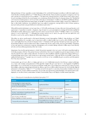
Table 3: Clinical pearls in identification of epithelial dystrophies 3,7,8,13,19
Feature Location Dystrophy
Map (geographic-shaped opacities)
Dot (putty-like opacities), Fingerprint Epithelial/diffuse EBMD
(whorl-like clustered lines)
Epithelial/concentrated in the interpalpebral
Epithelial vesicle of uniform shape and size Meesmann
region
Feathery and/or flame-shaped opacities and Epithelial/diffuse Lisch
optically empty microcysts
Fine reticular opacification Subepithelial/most dense in central or mid- Reis-Bücklers
(linear, ring-like, or alveolar patterns) periphery (extreme periphery is spared)
Subepithelial/most dense in central or mid-
Honeycomb patterned opacity Thiel-Behnke
periphery (extreme periphery is spared)
CANADIAN JOURNAL of OPTOMETRY | REVUE CANADIENNE D’OPTOMÉTRIE VOL. 79 NO. 4 15