
Page 13 - CJO_W17
P. 13
CASE STUDY
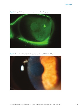
Figure 2: Topographical map corneal opacities present centrally in the left eye
Figure 3: Fluorescein staining highlights the geographic patterns of EBMD in the left eye
CANADIAN JOURNAL of OPTOMETRY | REVUE CANADIENNE D’OPTOMÉTRIE VOL. 79 NO. 4 13