
Page 37 - Gastrointestinal Bleeding (Xuất huyết tiêu hóa)
P. 37
CHAPTER 20 Gastrointestinal Bleeding 311
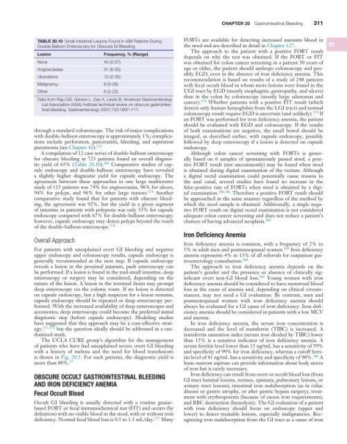
TABLE 20.10 Small Intestinal Lesions Found in 488 Patients During FOBTs are available for detecting increased amounts blood in
Double-Balloon Enteroscopy for Obscure GI Bleeding the stool and are described in detail in Chapter 127. 20
The approach to the patient with a positive FOBT result
Lesion Frequency, % (Range)
depends on why the test was obtained. If the FOBT or FIT
None 40 (0-57) was obtained for colon cancer screening in a patient 50 years of
Angioectasias 31 (6-55) age or older, the patient should undergo colonoscopy and pos-
sibly EGD, even in the absence of iron deficiency anemia. This
Ulcerations 13 (2-35) recommendation is based on results of a study of 248 patients
Malignancy 8 (3-26) with fecal occult blood in whom more lesions were found in the
Other 6 (2-22) UGI tract by EGD (mostly esophagitis, gastropathy, and ulcers)
than in the colon by colonoscopy (mostly large adenomas and
Data from Raju GS, Gerson L, Das A, Lewis B. American Gastroenterolog- cancer). 378 Whether patients with a positive FIT result (which
ical Association (AGA) Institute technical review on obscure gastrointes-
tinal bleeding. Gastroenterology 2007;133:1697–717. detects only human hemoglobin from the LGI tract) and normal
colonoscopy result require EGD is uncertain (and unlikely). 379 If
an FOBT was performed for iron deficiency anemia, the patient
should be evaluated with EGD and colonoscopy. If the results
through a standard colonoscope. The risk of major complications of both examinations are negative, the small bowel should be
with double-balloon enteroscopy is approximately 1%; complica- imaged, as described earlier, with capsule endoscopy, possibly
tions include perforation, pancreatitis, bleeding, and aspiration followed by deep enteroscopy if a lesion is detected on capsule
pneumonia (see Chapter 42). 373 endoscopy.
A compilation of 12 case series of double-balloon enteroscopy Although colon cancer screening with FOBTs is gener-
for obscure bleeding in 723 patients found an overall diagnos- ally based on 6 samples of spontaneously passed stool, a posi-
tic yield of 65% (Table 20.10). 309 Comparative studies of cap- tive FOBT result (not uncommonly) may be found when stool
sule endoscopy and double-balloon enteroscopy have revealed is obtained during digital examination of the rectum. Although
a slightly higher diagnostic yield for capsule endoscopy. The a digital rectal examination could potentially cause trauma to
agreement between these approaches in one large multicenter the anal canal, several studies have found no increase in the
study of 115 patients was 74% for angioectasias, 96% for ulcers, false-positive rate of FOBTs when stool is obtained by a digi-
94% for polyps, and 96% for other large tumors. 374 Another tal examination. 380,381 Therefore a positive FOBT result should
comparative study found that for patients with obscure bleed- be approached in the same manner regardless of the method by
ing, the agreement was 92%, but the yield in a given segment which the stool sample is obtained. Additionally, a single nega-
of intestine in patients with polyposis was only 33% for capsule tive FOBT result on digital rectal examination is not considered
endoscopy compared with 67% for double-balloon enteroscopy; adequate colon cancer screening and does not reduce a patient’s
however, capsule endoscopy may detect polyps beyond the reach chances of having advanced neoplasia. 382
of the double-balloon enteroscope. 374
Iron Deficiency Anemia
Overall Approach
Iron deficiency anemia is common, with a frequency of 2% to
For patients with unexplained overt GI bleeding and negative 5% in adult men and postmenopausal women. 383 Iron deficiency
upper endoscopy and colonoscopy results, capsule endoscopy is anemia represents 4% to 13% of all referrals for outpatient gas-
generally recommended as the next step. If capsule endoscopy troenterology consultation. 384
reveals a lesion in the proximal jejunum, push enteroscopy can The approach to iron deficiency anemia depends on the
be performed. If a lesion is found in the mid-small intestine, deep patient’s gender and the presence or absence of clinically sig-
enteroscopy or surgery may be considered, depending on the nificant overt non-GI blood loss. 385 Young women with iron
nature of the lesion. A lesion in the terminal ileum may prompt deficiency anemia should be considered to have menstrual blood
deep enteroscopy via the colonic route. If no lesion is detected loss as the cause of anemia and, depending on clinical circum-
on capsule endoscopy, but a high suspicion for a lesion remains, stances, may not need a GI evaluation. By contrast, men and
capsule endoscopy should be repeated or deep enteroscopy per- postmenopausal women with iron deficiency anemia should
formed. With the increased availability of deep enteroscopes and always be evaluated for a GI cause of iron deficiency. Iron defi-
accessories, deep enteroscopy could become the preferred initial ciency anemia should be considered in patients with a low MCV
diagnostic step (before capsule endoscopy). Modeling studies and anemia.
have suggested that this approach may be a cost-effective strat- In iron deficiency anemia, the serum iron concentration is
egy, 375,376 but the question ideally should be addressed in a ran- decreased and the level of transferrin (TIBC) is increased. A
domized study. transferrin saturation index (serum iron divided by TIBC) lower
The UCLA CURE group’s algorithm for the management than 15% is a sensitive indicator of iron deficiency anemia. A
of patients who have had unexplained severe overt GI bleeding serum ferritin level lower than 15 ng/mL has a sensitivity of 59%
with a history of melena and the need for blood transfusions and specificity of 99% for iron deficiency, whereas a cutoff ferri-
is shown in Fig. 20.5. For such patients, the diagnostic yield is tin level of 41 ng/mL has a sensitivity and specificity of 98%. 386 A
57
more than 80%. bone marrow aspirate can provide information about body stores
of iron but is rarely necessary.
Iron deficiency can result from overt or occult blood loss (from
OBSCURE OCCULT GASTROINTESTINAL BLEEDING GI tract luminal lesions, menses, epistaxis, pulmonary lesions, or
AND IRON DEFICIENCY ANEMIA urinary tract lesions), intestinal iron malabsorption (as in celiac
Fecal Occult Blood disease or gastric atrophy, or after gastric bypass surgery), treat-
ment with erythropoietin (because of excess iron requirements),
Occult GI bleeding is usually detected with a routine guaiac- and RBC destruction (hemolysis). The GI evaluation of a patient
based FOBT or fecal immunochemical test (FIT) and occurs (by with iron deficiency should focus on endoscopy (upper and
definition) with no visible blood in the stool, with or without iron lower) to detect treatable lesions, especially malignancies. Rec-
deficiency. Normal fecal blood loss is 0.5 to 1.5 mL/day. 377 Many ognizing iron malabsorption from the GI tract as a cause of iron