
Page 32 - Gastrointestinal Bleeding (Xuất huyết tiêu hóa)
P. 32
306 PART III Symptoms, Signs, and Biopsychosocial Issues
BOX 20�2 Causes of Obscure GI Bleeding
UPPER GI TRACT
Cameron lesions
Dieulafoy lesions
GAVE
SMALL INTESTINE
Angioectasia
Aortoenteric fistula
Dieulafoy lesion
Diverticulosis
Meckel diverticulum
Neoplasm
Pancreatic or biliary disease
Ulceration
COLON
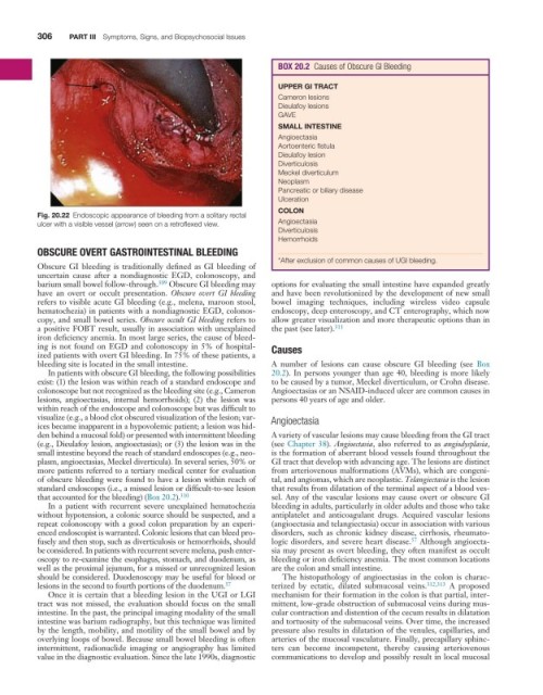
Fig. 20.22 Endoscopic appearance of bleeding from a solitary rectal
ulcer with a visible vessel (arrow) seen on a retroflexed view. Angioectasia
Diverticulosis
Hemorrhoids
OBSCURE OVERT GASTROINTESTINAL BLEEDING
*After exclusion of common causes of UGI bleeding.
Obscure GI bleeding is traditionally defined as GI bleeding of
uncertain cause after a nondiagnostic EGD, colonoscopy, and
barium small bowel follow-through. 309 Obscure GI bleeding may options for evaluating the small intestine have expanded greatly
have an overt or occult presentation. Obscure overt GI bleeding and have been revolutionized by the development of new small
refers to visible acute GI bleeding (e.g., melena, maroon stool, bowel imaging techniques, including wireless video capsule
hematochezia) in patients with a nondiagnostic EGD, colonos- endoscopy, deep enteroscopy, and CT enterography, which now
copy, and small bowel series. Obscure occult GI bleeding refers to allow greater visualization and more therapeutic options than in
a positive FOBT result, usually in association with unexplained the past (see later). 311
iron deficiency anemia. In most large series, the cause of bleed-
ing is not found on EGD and colonoscopy in 5% of hospital- Causes
ized patients with overt GI bleeding. In 75% of these patients, a
bleeding site is located in the small intestine. A number of lesions can cause obscure GI bleeding (see Box
In patients with obscure GI bleeding, the following possibilities 20.2). In persons younger than age 40, bleeding is more likely
exist: (1) the lesion was within reach of a standard endoscope and to be caused by a tumor, Meckel diverticulum, or Crohn disease.
colonoscope but not recognized as the bleeding site (e.g., Cameron Angioectasias or an NSAID-induced ulcer are common causes in
lesions, angioectasias, internal hemorrhoids); (2) the lesion was persons 40 years of age and older.
within reach of the endoscope and colonoscope but was difficult to
visualize (e.g., a blood clot obscured visualization of the lesion; var- Angioectasia
ices became inapparent in a hypovolemic patient; a lesion was hid-
den behind a mucosal fold) or presented with intermittent bleeding A variety of vascular lesions may cause bleeding from the GI tract
(e.g., Dieulafoy lesion, angioectasias); or (3) the lesion was in the (see Chapter 38). Angioectasia, also referred to as angiodysplasia,
small intestine beyond the reach of standard endoscopes (e.g., neo- is the formation of aberrant blood vessels found throughout the
plasm, angioectasias, Meckel diverticula). In several series, 50% or GI tract that develop with advancing age. The lesions are distinct
more patients referred to a tertiary medical center for evaluation from arteriovenous malformations (AVMs), which are congeni-
of obscure bleeding were found to have a lesion within reach of tal, and angiomas, which are neoplastic. Telangiectasia is the lesion
standard endoscopes (i.e., a missed lesion or difficult-to-see lesion that results from dilatation of the terminal aspect of a blood ves-
that accounted for the bleeding) (Box 20.2). 310 sel. Any of the vascular lesions may cause overt or obscure GI
In a patient with recurrent severe unexplained hematochezia bleeding in adults, particularly in older adults and those who take
without hypotension, a colonic source should be suspected, and a antiplatelet and anticoagulant drugs. Acquired vascular lesions
repeat colonoscopy with a good colon preparation by an experi- (angioectasia and telangiectasia) occur in association with various
enced endoscopist is warranted. Colonic lesions that can bleed pro- disorders, such as chronic kidney disease, cirrhosis, rheumato-
57
fusely and then stop, such as diverticulosis or hemorrhoids, should logic disorders, and severe heart disease. Although angioecta-
be considered. In patients with recurrent severe melena, push enter- sia may present as overt bleeding, they often manifest as occult
oscopy to re-examine the esophagus, stomach, and duodenum, as bleeding or iron deficiency anemia. The most common locations
well as the proximal jejunum, for a missed or unrecognized lesion are the colon and small intestine.
should be considered. Duodenoscopy may be useful for blood or The histopathology of angioectasias in the colon is charac-
lesions in the second to fourth portions of the duodenum. 57 terized by ectatic, dilated submucosal veins. 312,313 A proposed
Once it is certain that a bleeding lesion in the UGI or LGI mechanism for their formation in the colon is that partial, inter-
tract was not missed, the evaluation should focus on the small mittent, low-grade obstruction of submucosal veins during mus-
intestine. In the past, the principal imaging modality of the small cular contraction and distention of the cecum results in dilatation
intestine was barium radiography, but this technique was limited and tortuosity of the submucosal veins. Over time, the increased
by the length, mobility, and motility of the small bowel and by pressure also results in dilatation of the venules, capillaries, and
overlying loops of bowel. Because small bowel bleeding is often arteries of the mucosal vasculature. Finally, precapillary sphinc-
intermittent, radionuclide imaging or angiography has limited ters can become incompetent, thereby causing arteriovenous
value in the diagnostic evaluation. Since the late 1990s, diagnostic communications to develop and possibly result in local mucosal