
Page 30 - Gastrointestinal Bleeding (Xuất huyết tiêu hóa)
P. 30
304 PART III Symptoms, Signs, and Biopsychosocial Issues
The colonoscopic appearance of the mucosa includes erythema,
friability, and exudate. Mucosal biopsy specimens may suggest
ischemic changes but are generally used to exclude infectious
or Crohn colitis. Ischemic colitis usually resolves in a few days
and generally does not require colonoscopic hemostasis or anti-
biotic therapy. In the UCLA CURE experience, approximately
10% of patients with ischemic colitis and severe hematochezia
had a focal ulcer with a major stigma of hemorrhage on urgent
colonoscopy. 288 After detection of arterial blood flow with DEP,
the recommended treatment in these cases is epinephrine injec-
tion and hemoclipping, similar to that for other ulcers. In a large
retrospective series from Kaiser, no episodes of rebleeding from
ischemic colitis occurred over a 4-year follow-up period. 235 On
the other hand, patients with large-vessel mesenteric ischemia
usually have worse outcomes, including higher rates of rebleed-
ing, perforation, surgery, and death.
IBD that involves the colon can rarely cause severe acute LGI
bleeding (see Chapter 115). In a case series from the Mayo Clinic,
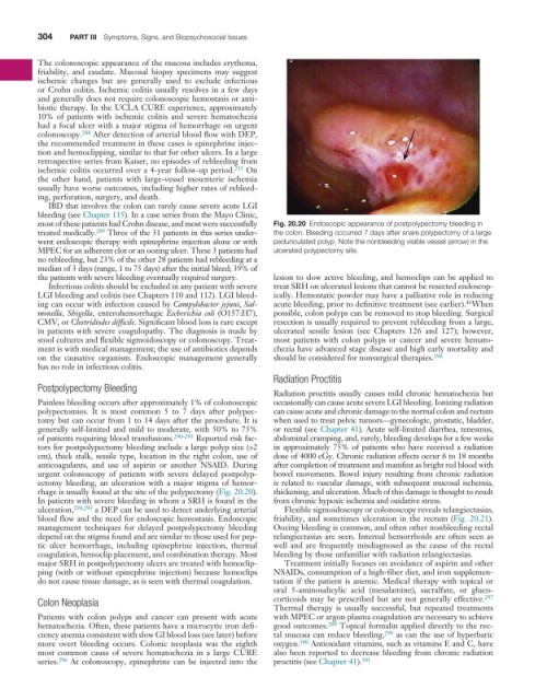
most of these patients had Crohn disease, and most were successfully Fig. 20.20 Endoscopic appearance of postpolypectomy bleeding in
treated medically. 289 Three of the 31 patients in this series under- the colon. Bleeding occurred 7 days after snare polypectomy of a large
went endoscopic therapy with epinephrine injection alone or with pedunculated polyp. Note the nonbleeding visible vessel (arrow) in the
MPEC for an adherent clot or an oozing ulcer. These 3 patients had ulcerated polypectomy site.
no rebleeding, but 23% of the other 28 patients had rebleeding at a
median of 3 days (range, 1 to 75 days) after the initial bleed; 39% of
the patients with severe bleeding eventually required surgery. lesion to slow active bleeding, and hemoclips can be applied to
Infectious colitis should be excluded in any patient with severe treat SRH on ulcerated lesions that cannot be resected endoscop-
LGI bleeding and colitis (see Chapters 110 and 112). LGI bleed- ically. Hemostatic powder may have a palliative role in reducing
43
ing can occur with infection caused by Campylobacter jejuni, Sal- acute bleeding, prior to definitive treatment (see earlier). When
monella, Shigella, enterohemorrhagic Escherichia coli (O157:H7), possible, colon polyps can be removed to stop bleeding. Surgical
CMV, or Clostridiodes difficile. Significant blood loss is rare except resection is usually required to prevent rebleeding from a large,
in patients with severe coagulopathy. The diagnosis is made by ulcerated sessile lesion (see Chapters 126 and 127); however,
stool cultures and flexible sigmoidoscopy or colonoscopy. Treat- most patients with colon polyps or cancer and severe hemato-
ment is with medical management; the use of antibiotics depends chezia have advanced stage disease and high early mortality and
on the causative organism. Endoscopic management generally should be considered for nonsurgical therapies. 296
has no role in infectious colitis.
Radiation Proctitis
Postpolypectomy Bleeding
Radiation proctitis usually causes mild chronic hematochezia but
Painless bleeding occurs after approximately 1% of colonoscopic occasionally can cause acute severe LGI bleeding. Ionizing radiation
polypectomies. It is most common 5 to 7 days after polypec- can cause acute and chronic damage to the normal colon and rectum
tomy but can occur from 1 to 14 days after the procedure. It is when used to treat pelvic tumors—gynecologic, prostatic, bladder,
generally self-limited and mild to moderate, with 50% to 75% or rectal (see Chapter 41). Acute self-limited diarrhea, tenesmus,
of patients requiring blood transfusions. 290-293 Reported risk fac- abdominal cramping, and, rarely, bleeding develops for a few weeks
tors for postpolypectomy bleeding include a large polyp size (>2 in approximately 75% of patients who have received a radiation
cm), thick stalk, sessile type, location in the right colon, use of dose of 4000 cGy. Chronic radiation effects occur 6 to 18 months
anticoagulants, and use of aspirin or another NSAID. During after completion of treatment and manifest as bright red blood with
urgent colonoscopy of patients with severe delayed postpolyp- bowel movements. Bowel injury resulting from chronic radiation
ectomy bleeding, an ulceration with a major stigma of hemor- is related to vascular damage, with subsequent mucosal ischemia,
rhage is usually found at the site of the polypectomy (Fig. 20.20). thickening, and ulceration. Much of this damage is thought to result
In patients with severe bleeding in whom a SRH is found in the from chronic hypoxic ischemia and oxidative stress.
ulceration, 294,295 a DEP can be used to detect underlying arterial Flexible sigmoidoscopy or colonoscopy reveals telangiectasias,
blood flow and the need for endoscopic hemostasis. Endoscopic friability, and sometimes ulceration in the rectum (Fig. 20.21).
management techniques for delayed postpolypectomy bleeding Oozing bleeding is common, and often other nonbleeding rectal
depend on the stigma found and are similar to those used for pep- telangiectasias are seen. Internal hemorrhoids are often seen as
tic ulcer hemorrhage, including epinephrine injection, thermal well and are frequently misdiagnosed as the cause of the rectal
coagulation, hemoclip placement, and combination therapy. Most bleeding by those unfamiliar with radiation telangiectasias.
major SRH in postpolypectomy ulcers are treated with hemoclip- Treatment initially focuses on avoidance of aspirin and other
ping (with or without epinephrine injection) because hemoclips NSAIDs, consumption of a high-fiber diet, and iron supplemen-
do not cause tissue damage, as is seen with thermal coagulation. tation if the patient is anemic. Medical therapy with topical or
oral 5-aminosalicylic acid (mesalamine), sucralfate, or gluco-
Colon Neoplasia corticoids may be prescribed but are not generally effective. 297
Thermal therapy is usually successful, but repeated treatments
Patients with colon polyps and cancer can present with acute with MPEC or argon plasma coagulation are necessary to achieve
hematochezia. Often, these patients have a microcytic iron defi- good outcomes. 289 Topical formalin applied directly to the rec-
ciency anemia consistent with slow GI blood loss (see later) before tal mucosa can reduce bleeding, 299 as can the use of hyperbaric
more overt bleeding occurs. Colonic neoplasia was the eighth oxygen. 300 Antioxidant vitamins, such as vitamins E and C, have
most common cause of severe hematochezia in a large CURE also been reported to decrease bleeding from chronic radiation
series. 296 At colonoscopy, epinephrine can be injected into the proctitis (see Chapter 41). 301 