
Page 189 - C:\Users\uromn\Videos\seyyedi pdf\
P. 189
S.A. Seyyedi et al. Photodiagnosis and Photodynamic Therapy 49 (2024) 104282
recurrence of RHL [23,24]. Application of PBM on patients with recur- The lesion size was determined by measuring the two main diameters
rent herpes simplex infection shown to decrease the recurrence fre- of the lesion in square millimeters using a transparent graph paper and
quency of herpes labialis [23]. Another study found significant graded in range of 0 to 3 (grade 0: no lesion, grade 1: 0.1 to 2.0 mm,
reduction of remission frequency in patients with recurrent herpes grade 2: 2–5 mm, and grade 3: larger than 5 mm). The pain intensity was
simplex infection [24]. On contrary, laser irradiation in a murine model evaluated based on the visual analogue scale (VAS), which 0 represented
did not affect stablished herpes simplex virus latency [25]. no pain and 10 represented the most severe pain ever experienced. At
Despite available body of evidence, the question of usefulness of PBM the first session, the lesion size before treatment and pain intensity
on herpes simplex virus infections treatment parameters (recurrence, before and right after treatment were evaluated by an oral medicine
symptoms severity, etc.) remains to be determined. In this study, we specialist.
aimed to evaluate the effect of PBM in the treatment of RHL. Our null Patients attended in follow-up sessions. In each follow-up the
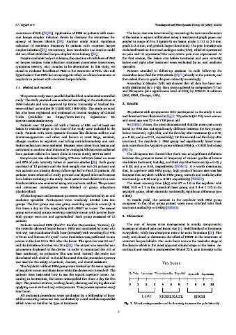
hypothesis is that PBM has no synergistic effect on clinical outcomes of researcher described the VAS criteria (Fig. 1) clearly to the patients, and
acyclovir in patients with recurrent herpes labialis then asked them to grade the pain intensity accordingly.
According to Shapiro Wilk test showed that all data has been nor-
1.1. Method and material mally distributed (p > 0.05). Data were analyzed by independent T-Test
and Chi-square (at a significance level of 0.05) by SPSS-21.0 software
The present study was a parallel double-blind randomized controlled (IBM, SPSS, Chicago, USA).
study. The study protocol was conducted according to the declaration of
1964-helsinki and was approved by Urmia University of Medical Sci- 2. Results
ences ethical committee IR.UMSU.REC.1400.048). The study protocol
has also been registered and evaluated at Iranian Registry of Clinical 22 patients with symptomatic RHL participated in the study A con-
Trials (available at: https://www.irct.ir/; registration ID: sort flowchart was illustrated in Fig. 2. 12 patients (54.4%) were women
IRCT20180224038840N2). and mean age was 27.5 ± 7.08 years old.
Patients over 18 years old with a history of RHL and at least one As Table 1 shows, the t-test demonstrated that the mean pain score
lesion in vesicular stage at the time of the study were included in the based on VAS was not significantly different between the two groups,
study. Patients with acute systemic diseases like diabetes mellitus and before treatment, right after, and the first day after treatment (p = 0.43,
immunosuppression and other oral lesions at crust stage, smokers, p = 0.55, and p = 0.12, respectively). On the second and third day after
allergic reaction to acyclovir and taking anti-inflammatory and anti- treatment, the Acyclovir + PBM group had significantly lower mean
biotic medications were excluded. Biopsies were taken from lesions and pain score than the Acyclovir group without PBM (p < 0.001 both days)
cultivated to confirm viral infection by virologist All data were collected (Fig. 3).
from patients referred to lasers clinic in Urmia during 2019 to 2021. The chi-square test showed that there was no significant difference
Sample size was calculated using G”Power software based on mean between the groups in terms of frequency of various grades of lesions
and STD of pain intensity values of previous studies [26]. Each group size before treatment, first day, and third day after treatment (p = 0.45, p
consisted of 12 patients and the final sample size was 24 patients and = 0.28, and p = 0.69, respectively) (Table 2). It is worth mentioning
two patients are missing during follow-ups led to final 22 patients. All that, in acyclovir with PBM group, high grade of lesions size was less
patients were informed of study protocol and signed informed consent frequent than acyclovir without PBM group, seventh and tenth day after
form before including in the study. Each patient was labeled with a code treatment (p = 0.03 and p = 0.001 respectively) (Table 2).
Randomization was conducted using toss and coin method. The patients The mean recovery time (day) was 8.7 ± 1.5 in the acyclovir whit
and outcomes investigators were blinded of group allocations PBM, 10.5 ± 1.1 in the turned-off laser group, and 3.4 ± 1.142 in the
(double-blind). acyclovir group, which showed a statistically significant difference (p =
All the diagnosis and treatment processes were performed by an oral 0.005).
medicine specialist. Participants were randomly divided into two As results yield, the patients in the acyclovir with PBM group
groups. The first group was case group receiving acyclovir cream (5 % compared to the other group patient were more satisfied whit their
five times a day for five days) along with PBMT as a case. The second treatment method (p = 0.008) (Table 3).
group was control group receiving acyclovir cream with passive laser.
Both groups were sex and age-matched. Each group consisted of 12 3. Discussion
patients.
Patients received PBM (Konf™- Konftec Corporation, Taiwan), (in The aim of herpes virus management is mainly symptomatic,
the vesicular phase of herpes lesions. PBM was conducted by mean of a focusing on clinical pain and lesion size [4]. Gold Standard of treatment
100 mW, semi-conductor diode laser (InGaAsP) with wavelength of 940 is Acyclovir, while low absorption rate is its main limitation [13]. This
2
±10 nm and fluence of 4 J/cm . Laser irradiation was performed in one study was aimed to determine the effect of PBMT in the treatment of
session in the first 24 to 48 h after the lesion. The spot size was 0.5 cm 2 recurrent herpes labialis. Our main focus was on the vesicular stage of
and the irritation duration was 20 s [23]. The output was recorded as a the disease which is the most apparent clinical stage of the lesion. Ac-
parameters displayed at the device. In order to minimized the risk of cording to our results in postoperative 48 and 72 h, pain intensity in the
laser scattering, no protective film was used. Instead, the probe was
disinfected with alcohol. It should be noted that the protective eyewear
was used for the safety of patients, dentists, and dental assistants.
The Acyclovir without PBM group were treated by the same regimen
of acyclovir cream and diode laser while the device was turned off. The
patients were instructed how to use the topical acyclovir cream. Ac-
cording to instructions, the cream was applied five times a day for five
days. The process involves, washing hands, cleaning and drying skin and
applying cream without any extra pressure. This process repeated every
four hours.
All treatment procedures were conducted by a fellowship of laser,
while measuring outcomes was conducted by a oral medicine specialist
which was not familiar by type of treatment. Fig. 1. Visual analogue scale used in the study to assess the pain intensity.
2