
Page 23 - ASOP ROT Study Guide
P. 23
It is probable that in cases of severe cartilage loss, the repair tissue is always by a combination of
collagen fibers and cartilage - fibrocartilage. The more the joint is kept moving, the greater will be
the ratio of cartilage to fibrous tissue. This is probably because, as outlined above, the
movement of a joint forces synovial fluid into the cartilage, supplying nutrients and removing
waste. Even in the most favorable circumstances pure cartilaginous replacement is not thought to
occur.
4 . 4 . 2 The Joint Capsule
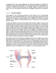
A joint capsule is a thin, fibrous sac containing fluid, which encloses a joint. The fluid provides
lubrication for bone movement. Most joints in the human body are freely movable and have much
more complex structures than the immovable or even the slightly movable types. The articular
(adjoining) ends of bones in a freely movable joint are covered with a thick layer of articular
cartilage, which is resistant to wear and produces a minimum of friction when it is compressed as
the joint is moved.
A tubular joint capsule that has two distinct layers holds joint bones together. The outer layer
consists mostly of dense, white, fibrous connective tissue, the fibers of which are attached to the
periosteum around the outside ring of each bone of the joint near its articular end. The outer fibrous
layer of the capsule, therefore, completely encloses the other parts of the joint. It is flexible enough,
though, to allow movement and strong enough to help prevent the articular surfaces from being
pulled apart. Bundles of strong, tough collagenous fibers called ligaments reinforce the joint
capsule and help to bind the articular ends of the bones together. Some ligaments appear as
bulges in the fibrous layer of the capsule, while others are accessory structures located outside the
capsule. In either case, these structures also prevent too much movement at the joint, because the
ligament is relatively inelastic and becomes tightly drawn whenever a normal limit of movement has
been achieved in the joint. The inner layer of the joint capsule consists of a shiny, vascular lining of
loose connective tissue called synovial membrane. The membrane covers all of the surfaces within
the joint capsule, except the areas that are covered by cartilage.
Some freely movable joints are partially or completely divided into two compartments by disks of
fibrocartilage called menisci located between the articular surfaces. Such a disk is attached to the
fibrous layer of the joint capsule at the sides, and its free surface projects into the joint cavity.
Certain freely movable joints also have closed, fluid-filled sacs called bursae associated with them.
Each bursa has an inner lining of synovial membrane, which may be continuous with the synovial
membrane of a nearby joint cavity. Bursae act as cushions and aid the movement of tendons,
which glide over such bony parts or over other tendons. The names of the bursae indicate their
locations; for example, a suprapatellar bursa, a prepatellar bursa, and an infrapatellar bursa.
4 . 5 Ligaments