
Page 51 - ASOP ROT Study Guide
P. 51
4. THE KNEE JOINT
4 . 1 Structure
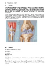
The knee joint is formed by the convex distal surfaces of the femur and the flat proximal surface of
the tibia (illustrated below). The knee cap (or patella) is a small sesamoid bone between the
muscular part of the quadriceps and the tendinous insertion of that muscle into the tibia. The patella
glides over the end of the femur during knee motion. The part of the synovial cavity of the knee in
which the patella moves is referred to, for descriptive convenience, as the patella-femoral
compartment.
The two convex femoral condyles move on the flat surface of the tibia. These two regions are
referred to, again for descriptive convenience, as the medial and lateral compartments. It is
important to realize that the medial, lateral and patella-femoral compartments of the knee are simply
regions of the same joint. The fibula does not form part of the knee joint except to act as an anchor
for knee muscles and ligaments.
www.webmd.com/pain-management/knee-pain/picture-of-the-knee
4 . 2 Stability
Four factors contribute to knee stability:
♦ Ligaments,
♦ Muscles,
♦ Menisci, and
♦ Shape
(Unlike the hip, the shape of the bones of the knee are such that they contribute very little to
stability.)
The stability of the knee joint stems mainly from its ligaments. On either side of the joint lie the
medial and lateral collateral ligaments (illustrated below). Within the joint are two large ligaments
which cross each other in the center of the knee joint. These are the anterior and posterior cruciate
ligaments.