
Page 169 - DUOKOPT BIBLIOBOOK
P. 169
EFFICACY
J Kwon, et al. Cytotoxic Effects of Dorzolamide/timolol
Viability analysis in the Cosopt-S group showed a uniform hexagonal ap-
The live/dead cell assay performed 24 hours after injec- pearance with regular cell borders and distinct microvilli
tion revealed that many endothelial cells in the Cosopt on the cell surface (Fig. 4B).
group were dead as evidenced by red-stained nuclei (Fig.
2C). However, in the Cosopt-S group, dead cells were rare-
ly observed (Fig. 2D). The median number of dead cells Discussion
from 5 consecutive microscopic fields (×400) on each eye
was 28 in the Cosopt group and 2 in the Cosopt-S group (p Ness et al. [15] recently reported that the Durasite bioad-
< 0.001) (Fig. 3A). hesive delivery system in topical antibiotics can block the
TUNEL staining demonstrated that distinct apoptosis of trabecular meshwork and have a toxic effect on rabbit cor-
endothelial cells was present only in the Cosopt group, and neal endothelial cells when introduced intracamerally. Im-
not the Cosopt-S group (Fig. 3B and 3C). The median mediately after cataract surgery or penetrating keratoplas-
number of TUNEL-positive endothelial cells, which were ty, topical eyedrops can penetrate into the anterior chamber
counted from 5 consecutive microscopic fields (×400), was through unstable wounds. Sutureless clear corneal surgery
4 in the Cosopt group and 0 in the Cosopt-S group (p < also can result in the tear film moving in and out of the eye
0.001) (Fig. 3A). during blinking if the wound leaks [11], which can be ex-
acerbated if the corneal epithelium is injured [16]. Under
such conditions, the concentration of eyedrops in the ante-
Scanning electron microscopy
rior chamber would be higher than in eyes with an intact
Under scanning electron microscopy, the corneal endo- epithelium.
thelium in the Cosopt group partially lost microvilli on the Fixed combination anti-glaucoma eyedrops have recent-
cell surface and the intercellular junctions were extensive- ly been introduced for better patient compliance and great-
ly destroyed (Fig. 4A). However, the corneal endothelium er intraocular pressure reduction. Cosopt is a fixed combi-
A B A B
C D C D
100 μm 100 μm 20 μm 20 μm
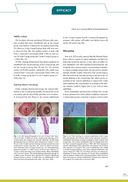
Fig. 1. (A) Slit lamp photograph of an eye 24 hours after injec- Fig. 2. (A) Vital staining of corneal endothelium with trypan
tion of Cosopt. A rabbit eye in the Cosopt group showing severe blue and alizarin red 24 hours after Cosopt injection. Extensive
corneal haze and conjunctival vascular injection. (B) Slit lamp endothelial cell damage is noted, resulting in nuclei stained with
photograph of an eye 24 hours after injection of Cosopt-S. A rab- trypan blue. The corneal endothelial cells are enlarged and have
bit eye in the Cosopt-S group showing minimal corneal haze and lost their normal hexagonal pattern (×400). (B) Vital staining of
conjunctival vascular injection, the extent of which was much corneal endothelium with trypan blue and alizarin red 24 hours
more mild than that of Cosopt-treated eyes. (C) Histopathologic after injection of Cosopt-S. The corneal endothelial cells exhibit
photomicrograph of a rabbit cornea 24 hours after injection of a normal hexagonal pattern, and some enlarged endothelial cells
Cosopt. Cornea showing severe stromal edema. Many endothelial can be observed (×400). (C) Live/dead cell assay on corneal en-
cells were lost (inset). (D) Histopathologic photomicrograph of dothelium 24 hours after injection of Cosopt. Many endothelial
a rabbit cornea 24 hours after injection of Cosopt-S. Significant cells are dead as evidenced by red-stained nuclei (×200). (D) Live/
stromal edema is absent. A single layer of endothelium is well dead cell assay on corneal endothelium 24 hours after injection
observed (inset) (hematoxylin and eosin, ×40; inset ×400). of Cosopt-S. Few dead cells are present (×200).
347
169