
Page 107 - Human Umbilical Cord Mesenchymal Stem Cells
P. 107
Chang, et al. / Tzu Chi Medical Journal 2018; 30(2): 71‑80
a
b
c d e
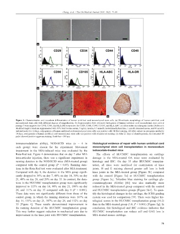
Figure 1: Characterization and mesoderm differentiation of human umbilical cord mesenchymal stem cells. (a) Fibroblastic morphology of human umbilical cord
mesenchymal stem cells with different degrees of magnification. (b) Representative flow cytometry histograms of human umbilical cord mesenchymal stem cells at
passage 3 were negative for CD34, CD45, and HLA-DR but positive for CD29, CD44, CD90, CD105, and HLA-ABC. (c) After culture in adipogenic media (Dulbecco’s
Modified Eagle’s Medium supplemented with 10% fetal bovine serum, 5 ug/mL insulin, 0.5 mmol/L isobutylmethylxanthine, 1 µmol/L dexamethasone, and 60 µmol/L
indomethacin) for 14 days, adipogenesis of human umbilical cord mesenchymal stem cells was positive with Oil Red staining. (d) After culture in osteogenic media for
14 days, osteogenesis of human umbilical cord mesenchymal stem cells was positive with Alizarin red staining. (e) After 21 days of chondrogenesis, the formatted 3D
pallet showed positive aggrecan staining. Scale bar = 100 µm
immunomodulation ability), NOD-SCID mice (n = 6 in Histological evidence of repair with human umbilical cord
each group) were chosen for the experiment. Movement mesenchymal stem cell transplantation in monosodium
impairment in the MIA-induced mice was evaluated by the iodoacetate-treated mice
Rota-Rod test. Figure 4 demonstrates that on day 7 after MIA The effects of HUCMSC transplantation on cartilage
intra-articular injection, there was a significant impairment in damage in the MIA-treated OA mice were evaluated by
running duration in the NOD/SCID mice (MIA-treated group) histology and IHC. On day 35 after HUCMSC transplan-
compared with the control group (P < 0.05). Running dura- tation, all mice were sacrificed for examination of knee
tions in the Rota-Rod test were evaluated after MIA-treatment. joints. H and E staining showed greater cell loss in both
Compared with day 0, the duration in the MIA group signifi- knee joints in the MIA-treated group [Figure 5b] compared
cantly dropped to 36% on day 7, 40% on day 14, 30% on day with the control [Figure 5a] or HUCMSC transplantation
21, 40% on day 28, and 20% on day 35. In contrast, the dura- group [Figure 5c]. Toluidine blue staining for cartilage gly-
tions in the HUCMSC transplantation group were significantly cosaminoglycans (GAGs) [41] was also markedly more
improved to 115% on day 14, 90% on day 21, 100% on day reduced in the MIA-treated group compared with the control
28, and 111% on day 35 compared with day 0 (P < 0.001). and HUCMSC transplantation groups [Figure 5d-f]. To quan-
These data were not significantly different from those of the tify the histological changes in the cartilage, the ICRS grading
control group, in which the running durations were 107% on system was used for comparison [39]. There were higher his-
day 14, 111% on day 21, 107% on day 28, and 112% on day tological scores in the HUCMSC transplantation group (10.2)
35 [Figure 4]. These results demonstrated improvement in than in the MIA-treated group (3.8, P < 0.001) [Figure 5g]. In
the running duration of the HUCMSC transplantation group. conclusion, this histological and IHC evidence indicates that
This may further suggest reduction in mechanical pain due to HUCMSC transplantation can reduce cell and GAG loss in
improvement in the knee joint with HUCMSC transplantation. MIA-treated mouse cartilage.
75