
Page 109 - Human Umbilical Cord Mesenchymal Stem Cells
P. 109
Chang, et al. / Tzu Chi Medical Journal 2018; 30(2): 71‑80
that HUCMSC transplantation could decrease MIA-induced Discussion
chondrocyte apoptosis in vivo. The present experiment demonstrated that HUCMSCs
fulfilled the criteria of MSCs and exhibited mesoderm differenti-
ation potential that can differentiate into adipocytes, osteocytes,
and chondrocytes; HUCMSC-CM assisted MIA-treated chon-
drocytes in recovering from impaired proliferation and increased
apoptosis and in reducing MIA-enhanced caspase 3 expression.
The in vivo experiment substantiated that impaired movements
in mice with MIA-induced cartilage destruction could be attenu-
ated by HUCMSC transplantation; the histological and IHC
evidence indicated that HUCMSC transplantation reduced cell
and GAG loss in MIA-treated mice; HUCMSC transplanta-
tion assisted MIA-treated mice in the regeneration of hyaline
cartilage and/or repair of cartilage damage and in ameliorating
cartilage apoptosis. Thus, HUCMSCs may be a feasible stem
cell source for treatment in OA cartilage repair.
HUCMSCs have attracted much attention as a potential
cell source for regenerative medicine, including OA [42]. The
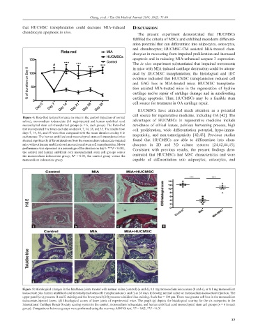
Figure 4: Rota-Rod test performance in mice in the control (injection of normal
saline), monosodium iodoacetate (0.1 mg)-injected and human umbilical cord advantages of HUCMSCs in regenerative medicine include
mesenchymal stem cell-transplanted groups (n = 6, each group). The Rota-Rod avoidance of ethical issues, painless harvesting process, high
test was repeated five times each day on days 0, 7, 14, 28, and 35. The results from cell proliferation, wide differentiation potential, hypo-immu-
days 7, 14, 28, and 35 were then compared with the mean duration on day 0 in nogenicity, and non-tumorigenicity [42,43]. Previous studies
each mouse. The human umbilical cord mesenchymal stem cell-transplanted mice
showed significantly different durations from the monosodium iodoacetate-injected found that HUCMSCs are able to differentiate into chon-
mice without human umbilical cord mesenchymal stem cell transplantation. Motor drocytes in 2D and 3D culture systems [24,42,44,45].
performance was expressed as a percentage of the duration on day 0. ***P < 0.001, Consistent with previous results, the present findings dem-
the control and human umbilical cord mesenchymal stem cell groups versus
the monosodium iodoacetate group; #P < 0.05, the control group versus the onstrated that HUCMSCs had MSC characteristics and were
monosodium iodoacetate group capable of differentiation into adipocytes, osteocytes, and
a b c g
d e f
Figure 5: Histological changes in the hind knee joints treated with normal saline (control) (a and d), 0.1 mg monosodium iodoacetate (b and e), or 0.1 mg monosodium
iodoacetate plus human umbilical cord mesenchymal stem cell transplantation (c and f) at 28 days following normal saline or monosodium iodoacetate injection. The
upper panel (a-c) presents H and E staining and the lower panel (d-f) presents toluidine blue staining. Scale bar = 100 µm. There was greater cell loss in the monosodium
iodoacetate-injected knees. (d) Histological scores of knee joints of experimental mice. The graph (g) depicts the histological scoring for the six categories in the
International Cartilage Repair Society scoring system in the control, monosodium iodoacetate, and human umbilical cord mesenchymal stem cell groups (n = 6 in each
group). Comparisons between groups were performed using the one-way ANOVA test. *P < 0.05, **P < 0.01
77