
Page 108 - Human Umbilical Cord Mesenchymal Stem Cells
P. 108
Chang, et al. / Tzu Chi Medical Journal 2018; 30(2): 71‑80
a
c b
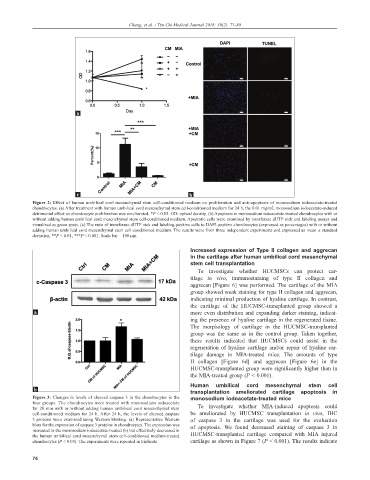
Figure 2: Effect of human umbilical cord mesenchymal stem cell-conditioned medium on proliferation and anti-apoptosis of monosodium iodoacetate-treated
chondrocytes. (a) After treatment with human umbilical cord mesenchymal stem cell-conditioned medium for 24 h, the 0.01 mg/mL monosodium iodoacetate-induced
detrimental effect on chondrocyte proliferation was ameliorated. *P < 0.05. OD: optical density. (b) Apoptosis in monosodium iodoacetate-treated chondrocytes with or
without adding human umbilical cord mesenchymal stem cell-conditioned medium. Apoptotic cells were examined by transferase dUTP nick end labeling assays and
visualized as green spots. (c) The ratio of transferase dUTP nick end labeling-positive cells to DAPI-positive chondrocytes (expressed as percentages) with or without
adding human umbilical cord mesenchymal stem cell-conditioned medium. The results were from three independent experiments and expressed as mean ± standard
deviation, **P < 0.01, ***P < 0.001. Scale bar = 100 µm
Increased expression of Type II collagen and aggrecan
in the cartilage after human umbilical cord mesenchymal
stem cell transplantation
To investigate whether HUCMSCs can protect car-
tilage in vivo, immunostaining of type II collagen and
aggrecan [Figure 6] was performed. The cartilage of the MIA
group showed weak staining for type II collagen and aggrecan,
indicating minimal production of hyaline cartilage. In contrast,
the cartilage of the HUCMSC-transplanted group showed a
a more even distribution and expanding darker staining, indicat-
ing the presence of hyaline cartilage in the regenerated tissue.
The morphology of cartilage in the HUCMSC-transplanted
group was the same as in the control group. Taken together,
these results indicated that HUCMSCs could assist in the
regeneration of hyaline cartilage and/or repair of hyaline car-
tilage damage in MIA-treated mice. The amounts of type
II collagen [Figure 6d] and aggrecan [Figure 6e] in the
HUCMSC-transplanted group were significantly higher than in
the MIA-treated group (P < 0.001).
Human umbilical cord mesenchymal stem cell
b transplantation ameliorated cartilage apoptosis in
Figure 3: Changes in levels of cleaved caspase 3 in the chondrocytes in the monosodium iodoacetate-treated mice
four groups. The chondrocytes were treated with monosodium iodoacetate
for 20 min with or without adding human umbilical cord mesenchymal stem To investigate whether MIA-induced apoptosis could
cell-conditioned medium for 24 h. After 24 h, the levels of cleaved caspase be ameliorated by HUCMSC transplantation in vivo, IHC
3 proteins were examined using Western blotting. (a) Representative Western of caspase 3 in the cartilage was used for the evaluation
blots for the expression of caspase 3 proteins in chondrocytes. The expression was of apoptosis. We found decreased staining of caspase 3 in
increased in the monosodium iodoacetate-treated (b) but effectively decreased in
the human umbilical cord mesenchymal stem cell-conditioned medium-treated HUCMSC-transplanted cartilage compared with MIA injured
chondrocytes (P < 0.05). The experiments were repeated in triplicate cartilage as shown in Figure 7 (P < 0.001). The results indicate
76