
Page 496 - Atlas of Small Animal CT and MRI
P. 496
486 Atlas of Small Animal CT and MRI
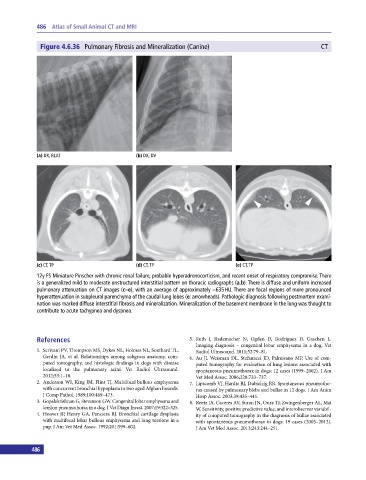
Figure 4.6.36 Pulmonary Fibrosis and Mineralization (Canine) CT
(a) DX, RLAT (b) DX, DV
(c) CT, TP (d) CT, TP (e) CT, TP
12y FS Miniature Pinscher with chronic renal failure, probable hyperadrenocorticism, and recent onset of respiratory compromise. There
is a generalized mild to moderate unstructured interstitial pattern on thoracic radiographs (a,b). There is diffuse and uniform increased
pulmonary attenuation on CT images (c–e), with an average of approximately −635 HU. There are focal regions of more pronounced
hyperattenuation in subpleural parenchyma of the caudal lung lobes (e: arrowheads). Pathologic diagnosis following postmortem exami-
nation was marked diffuse interstitial fibrosis and mineralization. Mineralization of the basement membrane in the lung was thought to
contribute to acute tachypnea and dyspnea.
References 5. Ruth J, Rademacher N, Ogden D, Rodriguez D, Gaschen L.
Imaging diagnosis – congenital lobar emphysema in a dog. Vet
1. Scrivani PV, Thompson MS, Dykes NL, Holmes NL, Southard TL, Radiol Ultrasound. 2011;52:79–81.
Gerdin JA, et al. Relationships among subgross anatomy, com- 6. Au JJ, Weisman DL, Stefanacci JD, Palmisano MP. Use of com-
puted tomography, and histologic findings in dogs with disease puted tomography for evaluation of lung lesions associated with
localized to the pulmonary acini. Vet Radiol Ultrasound. spontaneous pneumothorax in dogs: 12 cases (1999–2002). J Am
2012;53:1–10. Vet Med Assoc. 2006;228:733–737.
2. Anderson WI, King JM, Flint TJ. Multifocal bullous emphysema 7. Lipscomb VJ, Hardie RJ, Dubielzig RR. Spontaneous pneumotho-
with concurrent bronchial hypoplasia in two aged Afghan hounds. rax caused by pulmonary blebs and bullae in 12 dogs. J Am Anim
J Comp Pathol. 1989;100:469–473. Hosp Assoc. 2003;39:435–445.
3. Gopalakrishnan G, Stevenson GW. Congenital lobar emphysema and 8. Reetz JA, Caceres AV, Suran JN, Oura TJ, Zwingenberger AL, Mai
tension pneumothorax in a dog. J Vet Diagn Invest. 2007;19:322–325. W. Sensitivity, positive predictive value, and interobserver variabil-
4. Hoover JP, Henry GA, Panciera RJ. Bronchial cartilage dysplasia ity of computed tomography in the diagnosis of bullae associated
with multifocal lobar bullous emphysema and lung torsions in a with spontaneous pneumothorax in dogs: 19 cases (2003–2012).
pup. J Am Vet Med Assoc. 1992;201:599–602. J Am Vet Med Assoc. 2013;243:244–251.
486