
Page 492 - Atlas of Small Animal CT and MRI
P. 492
482 Atlas of Small Animal CT and MRI
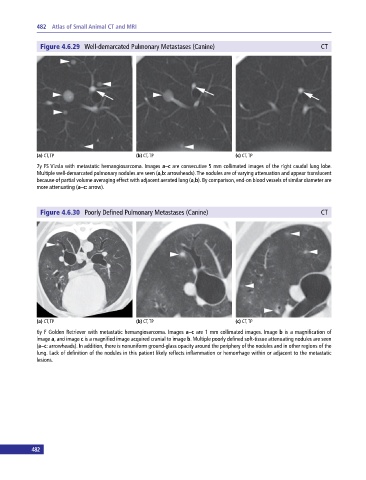
Figure 4.6.29 Well‐demarcated Pulmonary Metastases (Canine) CT
(a) CT, TP (b) CT, TP (c) CT, TP
7y FS Vizsla with metastatic hemangiosarcoma. Images a–c are consecutive 5 mm collimated images of the right caudal lung lobe.
Multiple well‐demarcated pulmonary nodules are seen (a,b: arrowheads). The nodules are of varying attenuation and appear translucent
because of partial volume averaging effect with adjacent aerated lung (a,b). By comparison, end‐on blood vessels of similar diameter are
more attenuating (a–c: arrow).
Figure 4.6.30 Poorly Defined Pulmonary Metastases (Canine) CT
(a) CT, TP (b) CT, TP (c) CT, TP
6y F Golden Retriever with metastatic hemangiosarcoma. Images a–c are 1 mm collimated images. Image b is a magnification of
image a, and image c is a magnified image acquired cranial to image b. Multiple poorly defined soft‐tissue attenuating nodules are seen
(a–c: arrowheads). In addition, there is nonuniform ground‐glass opacity around the periphery of the nodules and in other regions of the
lung. Lack of definition of the nodules in this patient likely reflects inflammation or hemorrhage within or adjacent to the metastatic
lesions.
482