
Page 487 - Atlas of Small Animal CT and MRI
P. 487
Small Airways and Parenchyma 477
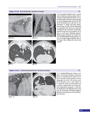
Figure 4.6.22 Bronchoalveolar Carcinoma (Canine) CT
11y M Australian Shepherd with no clinical
signs referable to pulmonary disease. There is
a large well‐demarcated soft‐tissue mass in
the dorsal aspect of the left caudal lung lobe
on survey radiographs (a,b: arrows). The soft‐
tissue attenuating mass is also seen on rep-
resentative CT images (c,d: large arrows).
Some airways are left relatively undisturbed
(c: arrowhead), while others are compressed
and displaced (d: arrowheads). Ground‐glass
opacities are present at the periphery of the
mass (c: small arrow). Pathologic diagnosis
following lung lobectomy was locally inva-
sive well‐differentiated bronchoalveolar car-
(a) DX, RLAT (b) DX, DV cinoma. The ground‐glass infiltrates seen on
CT images likely reflected the local tumor
invasion.
(c) CT, TP (d) CT, TP
Figure 4.6.23 Cavitary Bronchoalveolar Carcinoma (Canine) CT
11y FS Shepherd/Doberman Pinscher cross
with no clinical signs referable to pulmonary
disease. A well‐demarcated cavitary mass is
seen in the right caudal lung lobe on survey
radiographs (a: arrow). The cavitary nature
of the pulmonary mass is also evident on
CT images (b: arrow), and the presence of
a meniscus indicates that part of the soft‐
tissue attenuating component is fluid that
has distributed in the dependent part of the
mass (b: arrowhead). Pathologic diagnosis
following lung lobectomy was bronchoalve-
olar carcinoma.
(a) DX, LLAT (b) CT, TP
477