
Page 490 - Atlas of Small Animal CT and MRI
P. 490
480 Atlas of Small Animal CT and MRI
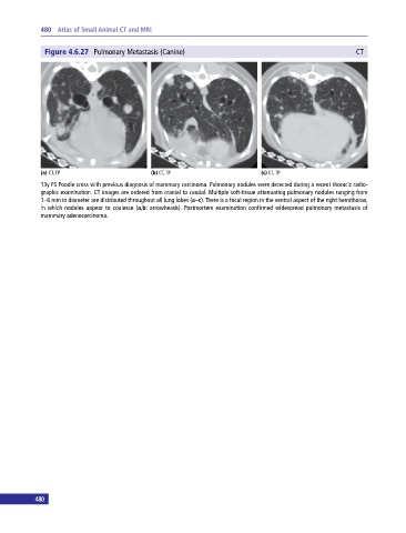
Figure 4.6.27 Pulmonary Metastasis (Canine) CT
(a) CT, TP (b) CT, TP (c) CT, TP
13y FS Poodle cross with previous diagnosis of mammary carcinoma. Pulmonary nodules were detected during a recent thoracic radio-
graphic examination. CT images are ordered from cranial to caudal. Multiple soft‐tissue attenuating pulmonary nodules ranging from
1–6 mm in diameter are distributed throughout all lung lobes (a–c). There is a focal region in the ventral aspect of the right hemithorax,
in which nodules appear to coalesce (a,b: arrowheads). Postmortem examination confirmed widespread pulmonary metastasis of
mammary adenocarcinoma.
480