
Page 489 - Atlas of Small Animal CT and MRI
P. 489
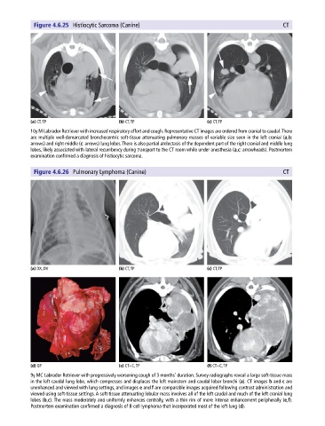
Figure 4.6.25  Histiocytic Sarcoma (Canine)                      Small Airways and Parenchyma  479
                                                                                                          CT


            (a) CT, TP                       (b) CT, TP                       (c) CT, TP
            10y M Labrador Retriever with increased respiratory effort and cough. Representative CT images are ordered from cranial to caudal. There
            are multiple well‐demarcated bronchocentric soft‐tissue attenuating pulmonary masses of variable size seen in the left cranial (a,b:
            arrows) and right middle (c: arrows) lung lobes. There is also partial atelectasis of the dependent part of the right cranial and middle lung
            lobes, likely associated with lateral recumbency during transport to the CT room while under anesthesia (a,c: arrowheads). Postmortem
            examination confirmed a diagnosis of histiocytic sarcoma.

              Figure 4.6.26  Pulmonary Lymphoma (Canine)                                                  CT


            (a) DX, DV                       (b) CT, TP                       (c) CT, TP


            (d) GP                           (e) CT+C, TP                     (f) CT+C, TP
            9y MC Labrador Retriever with progressively worsening cough of 3 months’ duration. Survey radiographs reveal a large soft‐tissue mass
            in the left caudal lung lobe, which compresses and displaces the left mainstem and caudal lobar bronchi (a). CT images b and c are
            unenhanced and viewed with lung settings, and images e and f are comparable images acquired following contrast administration and
            viewed using soft‐tissue settings. A soft‐tissue attenuating lobular mass involves all of the left caudal and much of the left cranial lung
            lobes (b,c). The mass moderately and uniformly enhances centrally, with a thin rim of more intense enhancement peripherally (e,f).
            Postmortem examination confirmed a diagnosis of B‐cell lymphoma that incorporated most of the left lung (d).