
Page 495 - Atlas of Small Animal CT and MRI
P. 495
Small Airways and Parenchyma 485
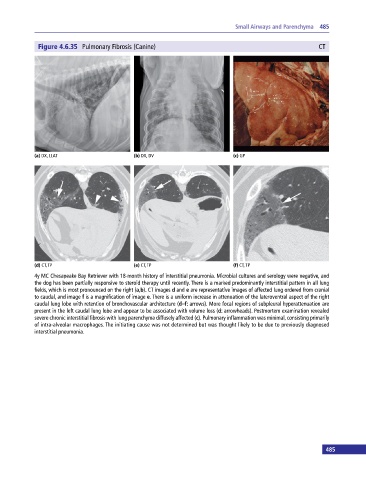
Figure 4.6.35 Pulmonary Fibrosis (Canine) CT
(a) DX, LLAT (b) DX, DV (c) GP
(d) CT, TP (e) CT, TP (f) CT, TP
4y MC Chesapeake Bay Retriever with 18‐month history of interstitial pneumonia. Microbial cultures and serology were negative, and
the dog has been partially responsive to steroid therapy until recently. There is a marked predominantly interstitial pattern in all lung
fields, which is most pronounced on the right (a,b). CT images d and e are representative images of affected lung ordered from cranial
to caudal, and image f is a magnification of image e. There is a uniform increase in attenuation of the lateroventral aspect of the right
caudal lung lobe with retention of bronchovascular architecture (d–f: arrows). More focal regions of subpleural hyperattenuation are
present in the left caudal lung lobe and appear to be associated with volume loss (d: arrowheads). Postmortem examination revealed
severe chronic interstitial fibrosis with lung parenchyma diffusely affected (c). Pulmonary inflammation was minimal, consisting primarily
of intra‐alveolar macrophages. The initiating cause was not determined but was thought likely to be due to previously diagnosed
interstitial pneumonia.
485