Page 70 - Atlas of Small Animal CT and MRI
P. 70

60  Atlas of Small Animal CT and MRI


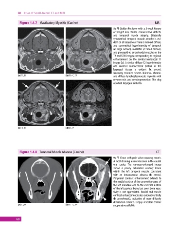
              Figure 1.4.7  Masticatory Myositis (Canine)                                                 MR
                                                                              8y FS Golden Retriever with a 2‐week history
                                                                              of  weight  loss,  stridor,  cranial  nerve  deficits,
                                                                              and temporal muscle atrophy. Bilaterally
                                                                                symmetrical temporal muscle atrophy is evi-
                                                                              dent on all sequences. There is marked, diffuse,
                                                                              and symmetrical hyperintensity of temporal
                                                                              (c: large arrows), masseter (c: small arrows),
                                                                              and pterygoid (c: arrowheads) muscles on the
                                                                              T2 and STIR images corresponding to regional
                                                                              enhancement on the contrast‐enhanced  T1
                                                                              image (b). A similar diffuse T2 hyperintensity
                                                                              and contrast enhancement pattern of the
                                                                                laryngeal tissues is evident (b: arrow).
                                                                              Necropsy revealed severe, bilateral, chronic,
             (a) T1, TP                       (b) T1+C, TP                    and diffuse lymphoplasmacytic myositis with
                                                                                myonecrosis and myodegeneration. This dog
                                                                              also had laryngeal cellulitis.


             (c) T2, TP                       (d) ST, TP


              Figure 1.4.8  Temporal Muscle Abscess (Canine)                                               CT

                                                                              9y FS Chow with pain when opening mouth.
                                                                              A focal draining lesion was seen in the caudal
                                                                              oral cavity.  The contrast‐enhanced image
                                                                              shows  a poorly delineated  cavitary lesion
                                                                              within the left temporal muscle, consistent
                                                                              with an intramuscular abscess (b: arrow).
                                                                              Peripheral  contrast  enhancement  extends  to
                                                                              the medial surface of the coronoid process of
                                                                              the left mandible and to the external surface
                                                                              of the left parietal bone, but overt bone reac-
                                                                              tivity is not appreciated. Fascial and muscle
                                                                              contrast enhancement is also evident ventrally
                                                                              (b:  arrowheads), indicative of more diffusely
                                                                              distributed  cellulitis.  Biopsy  revealed  chronic
             (a) CT, TP                       (b) CT+C, TP                    suppurative cellulitis.


            60
   65   66   67   68   69   70   71   72   73   74   75