
Page 74 - Atlas of Small Animal CT and MRI
P. 74
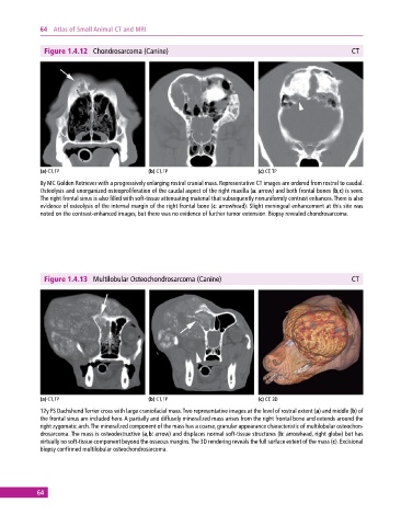
64  Atlas of Small Animal CT and MRI


              Figure 1.4.12  Chondrosarcoma (Canine)                                                       CT


             (a) CT, TP                       (b) CT, TP                       (c) CT, TP
             8y MC Golden Retriever with a progressively enlarging rostral cranial mass. Representative CT images are ordered from rostral to caudal.
             Osteolysis and unorganized osteoproliferation of the caudal aspect of the right maxilla (a: arrow) and both frontal bones (b,c) is seen.
             The right frontal sinus is also filled with soft‐tissue attenuating material that subsequently nonuniformly contrast enhances. There is also
             evidence of osteolysis of the internal margin of the right frontal bone (c: arrowhead). Slight meningeal enhancement at this site was
             noted on the contrast‐enhanced images, but there was no evidence of further tumor extension. Biopsy revealed chondrosarcoma.


              Figure 1.4.13  Multilobular Osteochondrosarcoma (Canine)                                     CT


             (a) CT, TP                       (b) CT, TP                       (c) CT, 3D
             12y FS Dachshund Terrier cross with large craniofacial mass. Two representative images at the level of rostral extent (a) and middle (b) of
             the frontal sinus are included here. A partially and diffusely mineralized mass arises from the right frontal bone and extends around the
             right zygomatic arch. The mineralized component of the mass has a coarse, granular appearance characteristic of multilobular osteochon-
             drosarcoma. The mass is osteodestructive (a,b: arrow) and displaces normal soft‐tissue structures (b: arrowhead, right globe) but has
             virtually no soft‐tissue component beyond the osseous margins. The 3D rendering reveals the full surface extent of the mass (c). Excisional
             biopsy confirmed multilobular osteochondrosarcoma.


            64