
Page 76 - Atlas of Small Animal CT and MRI
P. 76
66 Atlas of Small Animal CT and MRI
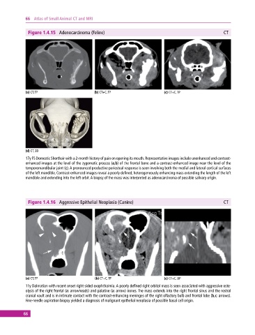
Figure 1.4.15 Adenocarcinoma (Feline) CT
(a) CT, TP (b) CT+C, TP (c) CT+C, TP
(d) CT, 3D
17y FS Domestic Shorthair with a 2‐month history of pain on opening its mouth. Representative images include unenhanced and contrast‐
enhanced images at the level of the zygomatic process (a,b) of the frontal bone and a contrast‐enhanced image near the level of the
temporomandibular joint (c). A pronounced productive periosteal response is seen involving both the medial and lateral cortical surfaces
of the left mandible. Contrast‐enhanced images reveal a poorly defined, heterogeneously enhancing mass extending the length of the left
mandible and extending into the left orbit. A biopsy of the mass was interpreted as adenocarcinoma of possible salivary origin.
Figure 1.4.16 Aggressive Epithelial Neoplasia (Canine) CT
(a) CT, TP (b) CT+C, TP (c) CT+C, DP
11y Dalmatian with recent onset right‐sided exophthalmia. A poorly defined right orbital mass is seen associated with aggressive oste-
olysis of the right frontal (a: arrowheads) and palatine (a: arrow) bones. The mass extends into the right frontal sinus and the rostral
cranial vault and is in intimate contact with the contrast‐enhancing meninges of the right olfactory bulb and frontal lobe (b,c: arrows).
Fine‐needle aspiration biopsy yielded a diagnosis of malignant epithelial neoplasia of possible basal cell origin.
66