
Page 73 - Atlas of Small Animal CT and MRI
P. 73
Skull 63
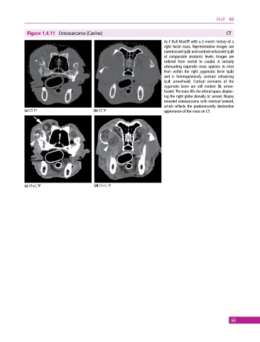
Figure 1.4.11 Osteosarcoma (Canine) CT
4y F Bull Mastiff with a 2‐month history of a
right facial mass. Representative images are
unenhanced (a,b) and contrast enhanced (c,d)
at comparable anatomic levels. Images are
ordered from rostral to caudal. A variably
attenuating expansile mass appears to arise
from within the right zygomatic bone (a,b)
and is heterogeneously contrast enhancing
(c,d: arrowhead). Cortical remnants of the
zygomatic bone are still evident (b: arrow-
heads). The mass fills the orbital space, displac-
ing the right globe dorsally (c: arrow). Biopsy
revealed osteosarcoma with minimal osteoid,
which reflects the predominantly destructive
(a) CT, TP (b) CT, TP appearance of the mass on CT.
(c) CT+C, TP (d) CT+C, TP
63