
Page 249 - Atlas of Small Animal CT and MRI
P. 249
Neoplasia 239
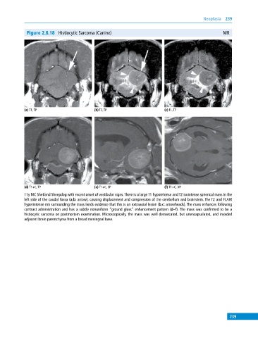
Figure 2.8.18 Histiocytic Sarcoma (Canine) MR
(a) T1, TP (b) T2, TP (c) FL, TP
(d) T1+C, TP (e) T1+C, SP (f) T1+C, DP
11y MC Shetland Sheepdog with recent onset of vestibular signs. There is a large T1 hypointense and T2 isointense spherical mass in the
left side of the caudal fossa (a,b: arrow), causing displacement and compression of the cerebellum and brainstem. The T2 and FLAIR
hyperintense rim surrounding the mass lends evidence that this is an extraaxial lesion (b,c: arrowheads). The mass enhances following
contrast administration and has a subtle nonuniform “ground glass” enhancement pattern (d–f). The mass was confirmed to be a
histiocytic sarcoma on postmortem examination. Microscopically, the mass was well demarcated, but unencapsulated, and invaded
adjacent brain parenchyma from a broad meningeal base.
239