
Page 257 - Atlas of Small Animal CT and MRI
P. 257
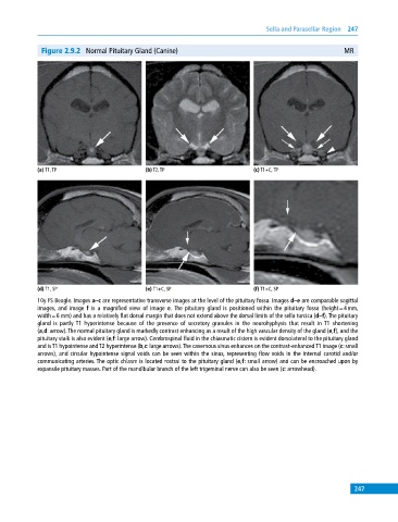
Sella and Parasellar Region  247


              Figure 2.9.2  Normal Pituitary Gland (Canine)                                              MR


            (a) T1, TP                       (b) T2, TP                       (c) T1+C, TP


            (d) T1, SP                       (e) T1+C, SP                     (f) T1+C, SP
            10y FS Beagle. Images a–c are representative transverse images at the level of the pituitary fossa. Images d–e are comparable sagittal
            images, and image  f is a magnified view of image  e. The pituitary gland is positioned within the pituitary fossa (height = 4 mm,
            width = 6 mm) and has a relatively flat dorsal margin that does not extend above the dorsal limits of the sella turcica (d–f). The pituitary
            gland is partly T1 hyperintense because of the presence of secretory granules in the neurohyphysis that result in T1 shortening
            (a,d: arrow). The normal pituitary gland is markedly contrast enhancing as a result of the high vascular density of the gland (e,f), and the
            pituitary stalk is also evident (e,f: large arrow). Cerebrospinal fluid in the chiasmatic cistern is evident dorsolateral to the pituitary gland
            and is T1  hypointense and T2 hyperintense (b,c: large arrows). The cavernous sinus enhances on the contrast‐enhanced T1 image (c: small
            arrows), and circular hypointense signal voids can be seen within the sinus, representing flow voids in the internal carotid and/or
              communicating arteries. The optic chiasm is located rostral to the pituitary gland (e,f: small arrow) and can be encroached upon by
            expansile pituitary masses. Part of the mandibular branch of the left trigeminal nerve can also be seen (c: arrowhead).


                                                                                                             247