
Page 258 - Atlas of Small Animal CT and MRI
P. 258
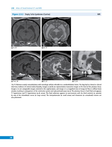
248  Atlas of Small Animal CT and MRI


              Figure 2.9.3  Empty Sella Syndrome (Canine)                                                 MR


             (a) T1, TP                       (b) T2, TP


             (c) T1+C, SP                     (d) T2, SP                       (e) T2, SP
             10y FS Pekinese acutely nonambulatory with neurologic deficits referable to a cerebrothalamic lesion. The dog had no clinical or clinical
             chemistry evidence of central endocrinopathy. Images a and b are representative transverse images at the level of the pituitary fossa.
             Images c–e are comparable images oriented in the sagittal plane, and image e is a magnified view of image d. There is diffuse brain
             atrophy resulting in enlargement of the ventricular system and subarachnoid space (a–e). The pituitary fossa is fluid filled and appears
             T1 hypointense and T2 hyperintense (a–d: arrow). This fluid collection appears to communicate with the third ventricle (e: asterisk)
             by way of the infundibular recess (e: large arrow). The interpeduncular (e: small arrow) and chiasmatic (e: arrowhead) cisterns are
             also prominent.


           248