
Page 444 - Atlas of Small Animal CT and MRI
P. 444
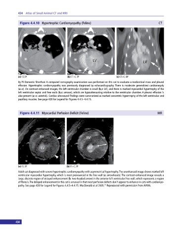
434  Atlas of Small Animal CT and MRI


              Figure 4.4.10  Hypertrophic Cardiomyopathy (Feline)                                          CT


             (a) CT, TP                       (b) CT+C, TP                     (c) CT+C, DP

             8y FS Domestic Shorthair. A computed tomography examination was performed on this cat to evaluate a mediastinal mass and pleural
             effusion. Hypertrophic cardiomyopathy was previously diagnosed by echocardiography. There is moderate generalized cardiomegaly
             (a–c). On contrast‐enhanced images, the left ventricular chamber is small (b,c: LV), and there is marked myocardial hypertrophy of the
             left ventricular septal and free walls (b,c: arrows), which are hypoattenuating relative to the ventricular chamber. A pleural effusion is
             also present (a–c: asterisk). Cardiac ultrasound findings were summarized as marked concentric hypertrophy of the left ventricular and
             papillary muscles. See page 428 for Legend for Figures 4.4.5–4.4.15.


              Figure 4.4.11  Myocardial Perfusion Deficit (Feline)                                        MR


             (a) T1, TP                       (b) T1+C, TP
             Adult cat diagnosed with severe hypertrophic cardiomyopathy with asymmetrical hypertrophy. The unenhanced image shows marked left
             ventricular myocardial hypertrophy, which is most pronounced in the free wall (a: arrowheads). The contrast‐enhanced image reveals a
             large, discrete region of delayed enhancement (b: two‐headed arrow) in the anterior left ventricular free wall, which represents a region
             of fibrosis. The delayed enhancement in this cat is unusual in that most perfusion defects don’t appear to enhance in cats with cardiomyo-
             pathy. See page 428 for Legend for Figures 4.4.5–4.4.15. MacDonald et al 2005.  Reproduced with permission from AVMA.
                                                                       19


           434