Page 445 - Atlas of Small Animal CT and MRI
P. 445

Heart, Pulmonary Vasculature, and Great Vessels  435


              Figure 4.4.12  Presumptive Right Atrial Hemangiosarcoma (Canine)                            CT
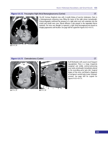
                                             10y MC German Shepherd cross with 3‐month history of exercise intolerance. There is
                                             a heterogeneously enhancing mass filling the lumen of the right atrium (arrowheads).
                                             Echocardiography confirmed the presence of a right atrial mass with extension into the
                                             cranial and caudal vena cava. Pleural effusion is also present in the dependent thorax
                                             (asterisk). The mass was thought to represent a right atrial hemangiosarcoma based on
                                             imaging appearance and location. See page 428 for Legend for Figures 4.4.5–4.4.15.


            (a) CT+C, TP


              Figure 4.4.13  Chemodectoma (Canine)                                                        CT

                                                                             9y M Rottweiler with recent onset frequent
                                                                             regurgitation. There is a large, irregularly
                                                                             margined and mildly contrast‐enhancing
                                                                             mass (a,b: arrowheads) immediately dorsal
                                                                             to the ascending aorta (a,b: AA). Surgical
                                                                             biopsy of the mass confirmed a diagnosis
                                                                             of malignant carotid body tumor (chemod-
                                                                             ectoma). See page 428 for Legend for
                                                                             Figures 4.4.5–4.4.15.


            (a) CT+C, TP                     (b) CT+C, SP


                                                                                                             435
   440   441   442   443   444   445   446   447   448   449   450