
Page 447 - Atlas of Small Animal CT and MRI
P. 447
Heart, Pulmonary Vasculature, and Great Vessels 437
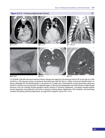
Figure 4.4.15 Pulmonary Hypertension (Canine) CT
(a) CT+C, TP (b) CT+C, TP (c) CT, MIP, DP
(d) DX, RLAT (e) CT, TP (f) CT, TP
11y FS Border Collie with rapid‐onset respiratory distress, lethargy, and inappetence. The pulmonary trunk (a: PT) and the right (a–c: RPA)
and left (a–c: LPA) pulmonary arteries are prominent. Descending aorta (a,b: DA). There is a diffuse unstructured interstitial pattern on
thoracic radiographs (d). The interstitial pattern is also present on CT images and is characterized by a patchy ground-glass appearance
(e,f) that is somewhat more pronounced in the dependent regions of the lung. Echocardiographic examination showed a mildly enlarged
pulmonary trunk and increased tricuspid regurgitant velocity, indicative of pulmonary hypertension. Lung biopsy revealed moderate
vascular hypertrophy and proliferation with acute to subacute multifocal alveolar degeneration, fibrin exudation, and hemorrhage,
consistent with primary pulmonary hypertension. See page 428 for Legend for Figures 4.4.5–4.4.15.
437