
Page 39 - Demo 1
P. 39
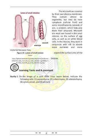
The microvilli are covered
                                                        by their own plasma membrane.
                                                        They    contain    almost    no
                                                        organelles,  but  they  do  have
                                                        cytoplasm  (cellular  fluid)  and
                                                        some microfilaments (strands of
                                                        acn, a protein), which help give
                                                        them  their  structure.  Microvilli
                                                        are most oen found in the small
                                                        intesne,  on  the  surface  of  egg
                                                        cells, as well as on white blood
                                                        cells. In the intesne, they work in
                                                        conjuncon  with  villi  to  absorb
                                                        more    nutrients   and   more
            material because they
                   Figure 24. Lumen of small intesne      expand the surface area of the
                              Source:

              https://upload.wikimedia.org/wikipedia/commons/thumb/  intesne.
              0/0e/Villi_%26_microvilli_of_small_intestine.svg/2000px-V
                  illi_%26_microvilli_of_small_intestine.svg.png


                     Learning Tasks and A ssesment


            Acvity 1. On   the  image  of  a  corn  stem  cross  secon  below,  indicate  the
                      following cells: (1) parenchyma, (2) collenchyma, (3) sclerenchyma,
                      (4) xylem vessel, and (5) phloem.


                                                 31