
Page 65 - Demo 1
P. 65
CELL MEMBRANE STRUCTURE: THE FLUID MOSAIC MODEL
The currently accepted model for the structure of the cell membrane,
called the Fluid Mosaic Model, was first proposed by the cell biologist S. J.
Singer and the biochemist G. L. Nicolson in 1972. This model has evolved over
me, but it sll provides a good basic descripon of the structure and behavior of
membranes in many cells.
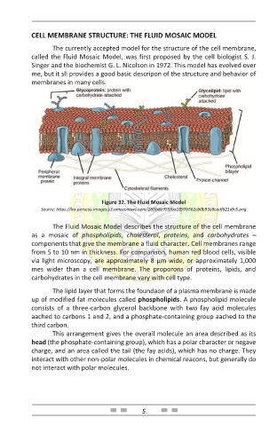
Figure 32. The Fluid Mosaic Model
Source: https://ka-perseus-images.s3.amazonaws.com/26ffb8e955fba1897b562cb0b93e8cadf621dfc5.png
The Fluid Mosaic Model describes the structure of the cell membrane
as a mosaic of phospholipids, cholesterol, proteins, and carbohydrates –
components that give the membrane a fluid character. Cell membranes range
from 5 to 10 nm in thickness. For comparison, human red blood cells, visible
via light microscopy, are approximately 8 µm wide, or approximately 1,000
mes wider than a cell membrane. The proporons of proteins, lipids, and
carbohydrates in the cell membrane vary with cell type.
The lipid layer that forms the foundaon of a plasma membrane is made
up of modified fat molecules called phospholipids. A phospholipid molecule
consists of a three-carbon glycerol backbone with two fay acid molecules
aached to carbons 1 and 2, and a phosphate-containing group aached to the
third carbon.
This arrangement gives the overall molecule an area described as its
head (the phosphate-containing group), which has a polar character or negave
charge, and an area called the tail (the fay acids), which has no charge. They
interact with other non-polar molecules in chemical reacons, but generally do
not interact with polar molecules.
57