
Page 191 - Clinical Manual of Small Animal Endosurgery
P. 191
Thoracoscopy 179
Endosurgical suturing and ligation
Extracorporeal suturing
Tying endosurgical locking slip knots or extracorporeal sutures, also
referred to as endoloops, formed outside the body, and then positioned
and tightened internally with a knot pusher, is an extremely useful tech-
nique to master in veterinary endosurgery. While commercially prepared
loops are available (Surgitie, Covidien; Endoloop, Ethicon) they cannot
be passed around fixed structures such as the ligamentum arteriosum,
plus it is more economical to prepare them oneself. Carpenter et al.
(2006) found hand-tied extracorporeal knots in both monofilament poly-
dioxanone and braided multifilament polyglactin 910 to be as secure and
reliable as commercially available endoloop ligatures for use in veteri-
nary endosurgery. They can be used to ligate vessels, including arteries
up to 3 mm in diameter, as well as for taking biopsies of lung and other
structures.
There are numerous different knots described that are suitable. The
initial endosurgical extracorporeal knot used was the Roeder knot (Hage,
2008). Originally implemented for tonsillectomies, its locking is unreli-
able unless used with catgut suture which swells on absorbing moisture,
‘locking’ the knot, and so it has fallen out of favour. There are several
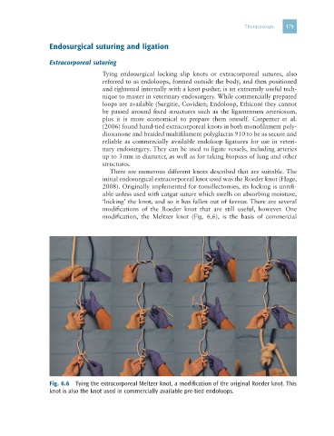
modifications of the Roeder knot that are still useful, however. One
modification, the Meltzer knot (Fig. 6.6), is the basis of commercial
Fig. 6.6 Tying the extracorporeal Meltzer knot, a modification of the original Roeder knot. This
knot is also the knot used in commercially available pre-tied endoloops.