
Page 196 - Clinical Manual of Small Animal Endosurgery
P. 196
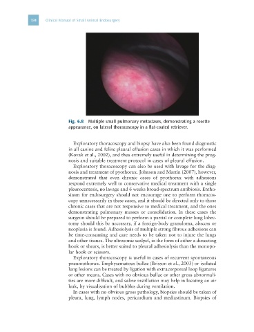
184   Clinical Manual of Small Animal Endosurgery


                              Fig. 6.8  Multiple small pulmonary metastases, demonstrating a rosette
                              appearance, on lateral thoracoscopy in a flat-coated retriever.


                                Exploratory thoracoscopy and biopsy have also been found diagnostic
                              in all canine and feline pleural effusion cases in which it was performed
                              (Kovak et al., 2002), and thus extremely useful in determining the prog-
                              nosis and suitable treatment protocol in cases of pleural effusion.
                                Exploratory thoracoscopy can also be used with lavage for the diag-
                              nosis and treatment of pyothorax. Johnson and Martin (2007), however,
                              demonstrated  that  even  chronic  cases  of  pyothorax  with  adhesions
                              respond extremely well to conservative medical treatment with a single
                              pleurocentesis, no lavage and 6 weeks broad-spectrum antibiosis. Enthu-
                              siasm for endosurgery should not encourage one to perform thoracos-
                              copy unnecessarily in these cases, and it should be directed only to those
                              chronic cases that are not responsive to medical treatment, and the ones
                              demonstrating  pulmonary  masses  or  consolidation.  In  these  cases  the
                              surgeon should be prepared to perform a partial or complete lung lobec-
                              tomy should this be necessary, if a foreign-body granuloma, abscess or
                              neoplasia is found. Adhesiolysis of multiple strong fibrous adhesions can
                              be time-consuming and care needs to be taken not to injure the lungs
                              and other tissues. The ultrasonic scalpel, in the form of either a dissecting
                              hook or shears, is better suited to pleural adhesiolysis than the monopo-
                              lar hook or scissors.
                                Exploratory thoracoscopy is useful in cases of recurrent spontaneous
                              pneumothorax. Emphysematous bullae (Brisson et al., 2003) or isolated
                              lung lesions can be treated by ligation with extracorporeal loop ligatures
                              or other means. Cases with no obvious bullae or other gross abnormali-
                              ties are more difficult, and saline instillation may help in locating an air
                              leak, by visualisation of bubbles during ventilation.
                                In cases with no obvious gross pathology, biopsies should be taken of
                              pleura,  lung,  lymph  nodes,  pericardium  and  mediastinum.  Biopsies  of