
Page 193 - Clinical Manual of Small Animal Endosurgery
P. 193
Thoracoscopy 181
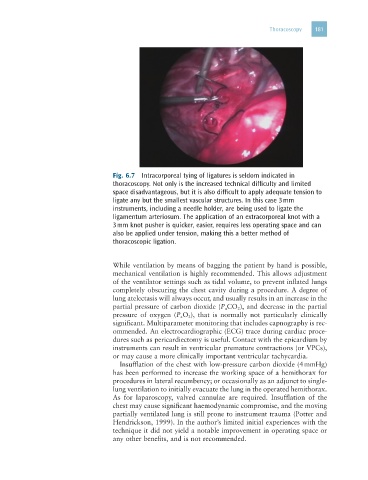
Fig. 6.7 Intracorporeal tying of ligatures is seldom indicated in
thoracoscopy. Not only is the increased technical difficulty and limited
space disadvantageous, but it is also difficult to apply adequate tension to
ligate any but the smallest vascular structures. In this case 3 mm
instruments, including a needle holder, are being used to ligate the
ligamentum arteriosum. The application of an extracorporeal knot with a
3 mm knot pusher is quicker, easier, requires less operating space and can
also be applied under tension, making this a better method of
thoracoscopic ligation.
While ventilation by means of bagging the patient by hand is possible,
mechanical ventilation is highly recommended. This allows adjustment
of the ventilator settings such as tidal volume, to prevent inflated lungs
completely obscuring the chest cavity during a procedure. A degree of
lung atelectasis will always occur, and usually results in an increase in the
partial pressure of carbon dioxide (P aCO 2), and decrease in the partial
pressure of oxygen (P a O 2 ), that is normally not particularly clinically
significant. Multiparameter monitoring that includes capnography is rec-
ommended. An electrocardiographic (ECG) trace during cardiac proce-
dures such as pericardiectomy is useful. Contact with the epicardium by
instruments can result in ventricular premature contractions (or VPCs),
or may cause a more clinically important ventricular tachycardia.
Insufflation of the chest with low-pressure carbon dioxide (4 mmHg)
has been performed to increase the working space of a hemithorax for
procedures in lateral recumbency; or occasionally as an adjunct to single-
lung ventilation to initially evacuate the lung in the operated hemithorax.
As for laparoscopy, valved cannulae are required. Insufflation of the
chest may cause significant haemodynamic compromise, and the moving
partially ventilated lung is still prone to instrument trauma (Potter and
Hendrickson, 1999). In the author’s limited initial experiences with the
technique it did not yield a notable improvement in operating space or
any other benefits, and is not recommended.